
At the 20-week anomaly scan, uterine artery Doppler can be used to look at the blood supply to the placenta. If you have been told that the resistance in your uterine arteries is increased, it is natural to have questions about what this means for you and your baby.
This guide explains what uterine artery Doppler measures, what the pulsatility index (PI) and notching mean, and what happens next if your result is abnormal.
Key points
- Uterine artery Doppler at 20 weeks assesses resistance to blood flow through the arteries supplying the uterus and placenta.
- An increased uterine artery PI or persistent notching may be associated with an increased likelihood of pre-eclampsia and fetal growth restriction.
- An abnormal result does not mean that the placenta is failing or that your baby will experience complications.
- Many women with increased uterine artery resistance have normal fetal growth and healthy pregnancies.
- The result is best interpreted alongside your risk factors, your baby's growth and other ultrasound findings, with additional monitoring where appropriate.
What is uterine artery Doppler?
Doppler is an ultrasound technique that measures how blood flows through a blood vessel. Uterine artery Doppler looks at the flow in the uterine arteries, the main blood vessels supplying the uterus and the placenta.
In a healthy pregnancy, blood flows easily to the placenta with low resistance. If the placenta has not developed normally, resistance in the uterine arteries rises, and this can be measured at the 20-week scan.
An increased result is a marker of increased risk, not a diagnosis. It does not mean that your baby will have complications.

How does blood flow to the placenta change during pregnancy?
During early pregnancy, the placenta establishes its blood supply through small maternal blood vessels known as the spiral arteries. As pregnancy progresses, cells from the developing placenta transform and remodel the spiral arteries into wider, low-resistance vessels that allow increasing amounts of maternal blood to reach the placenta.
This is known as spiral artery remodelling. As the placental circulation develops, resistance to blood flow through the uterine arteries therefore normally decreases.
In some pregnancies, however, these changes in the spiral arteries are incomplete, because the cells from the placenta do not fully remodel the arteries. This results in increased resistance to blood flow and can cause placental insufficiency, with reduced transfer of oxygen and nutrients from your circulation across the placenta to your baby. This can be associated with complications such as pre-eclampsia and fetal growth restriction.
Uterine artery Doppler provides an indirect assessment of these changes in the maternal placental circulation. It does not directly measure the amount of oxygen reaching your baby or establish whether the placenta is functioning normally.
Why is uterine artery Doppler performed at 20 weeks?
Uterine artery Doppler can be assessed at any stage of pregnancy. By 20–22 weeks, however, placental development is largely complete, so assessing blood flow in the uterine arteries at this stage shows whether the placental circulation has developed normally. Persistently increased resistance at this stage may be associated with an increased likelihood of:
- Pre-eclampsia.
- Fetal growth restriction.
- Having a baby who is small for gestational age.
- Certain complications associated with impaired placental function.
The examination can be particularly useful in women with additional risk factors, including a previous pregnancy affected by pre-eclampsia, fetal growth restriction or stillbirth.
How is uterine artery Doppler performed?
The examination can be performed transabdominally (through the tummy) or transvaginally (an internal scan). Colour Doppler ultrasound is used to identify the uterine arteries on either side of the uterus. A small sample of blood flow is recorded from each artery, producing a waveform that shows how blood flow velocity changes with each maternal heartbeat.
The pulsatility index is measured separately in the right and left uterine arteries. The average of these two measurements, known as the mean uterine artery PI, is generally used for clinical interpretation. The examination:
- Is non-invasive and does not require needles.
- Does not usually cause discomfort.
- Can be incorporated into the 20-week anomaly scan.
- Does not involve ionising radiation.
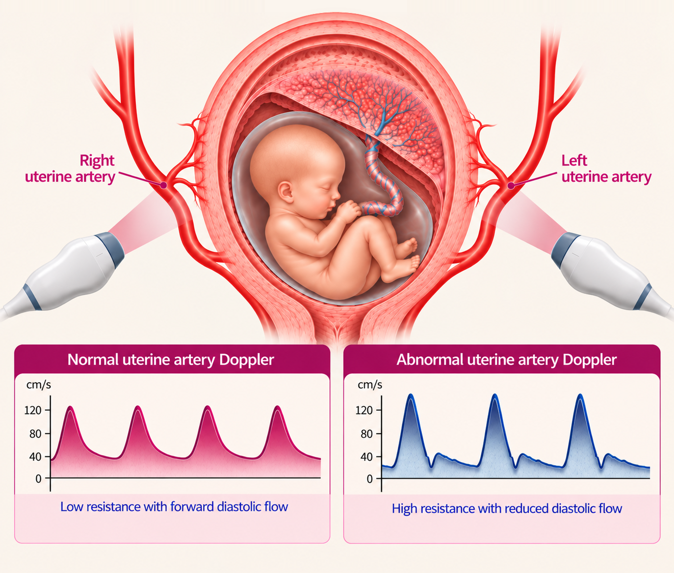
© The Pregnancy Clinic
The examination should be performed by appropriately trained professionals using recommended ultrasound safety settings.
What is uterine artery pulsatility index (PI)?
The pulsatility index, or PI, is the main measurement used to assess resistance to blood flow through the uterine arteries.
- A higher PI generally indicates greater resistance to blood flow.
- A lower PI generally indicates lower resistance.
During a normal pregnancy, uterine artery PI decreases as the maternal placental circulation develops. At approximately 20–22 weeks, an increased mean uterine artery PI is commonly defined as a measurement above the 95th centile for gestational age. This means that the resistance is higher than that seen in approximately 95% of pregnancies at a similar stage.
An increased PI indicates higher resistance to blood flow, but it does not mean that the placenta is failing.
What is uterine artery notching?
The uterine artery Doppler waveform may sometimes show a feature known as an early diastolic notch. During each heartbeat, blood flow velocity rises during systole, when the heart contracts, and decreases during diastole, when the heart relaxes. A notch is a small dip in the waveform at the start of diastole.
Notching is relatively common in early pregnancy because the placental circulation is still developing. As pregnancy progresses, notching becomes less frequent.
Persistent notching at 20–22 weeks, particularly when present in both uterine arteries, may be associated with increased resistance to blood flow and a greater likelihood of placental complications. However, it is a poor predictor of placental insufficiency, as many pregnancies with notching at this stage do not develop any complications.
For this reason, the mean uterine artery PI is generally the preferred measurement for assessing uterine artery resistance.
Is uterine artery Doppler at 20 weeks different from the 12-week examination?
Although the same arteries are examined, the clinical purpose differs.
At 12 weeks, uterine artery Doppler is primarily used as part of combined screening for pre-eclampsia. This screening may include maternal characteristics, mean arterial pressure, uterine artery PI and placental growth factor (PlGF) or pregnancy-associated plasma protein-A (PAPP-A). The aim is to identify women at increased risk of preterm pre-eclampsia who may benefit from preventive treatment with low-dose aspirin.
At 20–22 weeks, uterine artery Doppler provides information about resistance within the maternal placental circulation after placental vascular development is complete. The result may help identify women who would benefit from additional monitoring for fetal growth restriction and other placental complications.
| 12-week uterine artery Doppler | 20-week uterine artery Doppler | |
|---|---|---|
| Main purpose | Early screening for pre-eclampsia | Assessment of placental vascular resistance |
| Principal measurement | Mean uterine artery PI | Mean uterine artery PI |
| Clinical implications | Identifies women who may benefit from early preventive treatment | Helps determine the need for additional fetal growth surveillance |
| Important limitation | Cannot diagnose pre-eclampsia | Cannot on its own diagnose placental insufficiency or fetal growth restriction |
The two examinations are complementary but are not interchangeable.
For further information, read: Uterine Artery Doppler at 12 Weeks: What Is It and Why Is It Important?
Will I need extra growth scans if my uterine artery Doppler is abnormal?
Increased resistance in the uterine arteries at the 20–22-week anomaly scan is associated with the development of complications related to placental insufficiency, such as pre-eclampsia or fetal growth restriction, developing before 37 weeks. Uterine artery Doppler at this stage is poor at predicting late-onset complications that develop at term, i.e. after 37 weeks.
The chance of developing either of these complications is approximately 1 in 4 (25%), so the majority of pregnancies with an abnormal result do not develop them. The best way to monitor this is with:
- Regular fetal growth scans at 28, 32 and 36 weeks, to assess your baby's growth, with Doppler assessment of blood flow in the umbilical artery and your baby's brain (middle cerebral artery).
- Regular checks of your blood pressure.
How does The Pregnancy Clinic assess increased uterine artery resistance?
At The Pregnancy Clinic, we provide consultant-led ultrasound assessment for women with concerns about placental blood flow, fetal growth and pregnancy complications. Our approach combines specialist Doppler ultrasound with a detailed assessment of your individual pregnancy. This may include:
- Detailed review of the 20-week scan: assessment of fetal anatomy, growth and placental findings.
- Uterine artery Doppler: measurement of right and left uterine artery PI, interpreted using gestation-specific reference ranges.
- Individualised placental risk assessment: review of previous pregnancy complications, your medical history and first-trimester screening results.
- Specialist fetal growth assessment: ultrasound evaluation of fetal measurements, estimated fetal weight and growth centiles.
- Personalised pregnancy surveillance: recommendations for additional scans, coordinated with your NHS maternity team where appropriate.
Our aim is to provide clear explanations, appropriate reassurance and early identification of complications if they arise.
Related pregnancy guides
Frequently asked questions
What does increased uterine artery resistance at 20 weeks mean?
It means that blood flow through the arteries supplying the placenta meets more resistance than usual. It is a marker of increased risk of pre-eclampsia and fetal growth restriction, not a diagnosis, and many women with this result have healthy pregnancies.
What is a normal uterine artery PI at 20 weeks?
Uterine artery PI is interpreted against reference ranges for the stage of pregnancy. At 20–22 weeks, an increased mean PI is commonly defined as a value above the 95th centile for gestational age.
Is uterine artery notching a concern?
Persistent notching in both uterine arteries at 20–22 weeks may be associated with placental complications, but it is a poor predictor on its own. The mean uterine artery PI is the preferred measurement.
Will I need extra scans if my result is abnormal?
Usually, yes. Growth scans at about 28, 32 and 36 weeks, with Doppler assessment of the umbilical and middle cerebral arteries, together with regular blood pressure checks, are commonly recommended.
How is the 20-week test different from the 12-week test?
At 12 weeks, uterine artery Doppler is part of combined screening for pre-eclampsia, so that aspirin can be offered early. At 20–22 weeks, it assesses the placental circulation after its development is complete and helps decide whether extra growth monitoring is needed.
Specialist uterine artery Doppler assessment at The Pregnancy Clinic
At The Pregnancy Clinic, we offer consultant-led pregnancy ultrasound examinations, including uterine artery Doppler, detailed fetal growth assessment and individualised evaluation of placental function.
If you have been told that your uterine artery resistance is increased, or you have concerns about pre-eclampsia or fetal growth restriction, we can provide a detailed specialist assessment and explain what the findings mean for your pregnancy.
Book a specialist pregnancy ultrasound →This guide provides general information and does not replace advice from your maternity care team. The Pregnancy Clinic provides planned appointments only; for urgent concerns, please contact your maternity unit, NHS 111 or your GP.
Clinical references
- Papageorghiou AT, Yu CKH, Bindra R, Pandis G, Nicolaides KH. Multicenter screening for pre-eclampsia and fetal growth restriction by transvaginal uterine artery Doppler at 23 weeks of gestation. Ultrasound Obstet Gynecol. 2001;18:441–449.
- Akolekar R, Syngelaki A, Poon L, Wright D, Nicolaides KH. Competing risks model in early screening for preeclampsia by biophysical and biochemical markers. Fetal Diagn Ther. 2013;33:8–15.
- Rolnik DL, Wright D, Poon LC, et al. Aspirin versus placebo in pregnancies at high risk for preterm preeclampsia. N Engl J Med. 2017;377:613–622.
- Cnossen JS, Morris RK, ter Riet G, et al. Use of uterine artery Doppler ultrasonography to predict pre-eclampsia and intrauterine growth restriction: a systematic review and bivariable meta-analysis. CMAJ. 2008;178:701–711.
- National Institute for Health and Care Excellence (NICE). Hypertension in pregnancy: diagnosis and management. NICE guideline NG133.
- Fetal Medicine Foundation. Uterine artery Doppler assessment and screening for placental complications.
- NHS England. Saving Babies' Lives Care Bundle, Version 3. 2023.
- Royal College of Obstetricians and Gynaecologists (RCOG). Small-for-Gestational-Age Fetus and a Growth Restricted Fetus, Investigation and Care. Green-top Guideline No. 31. 2024.