
The placenta supports your baby's growth by transferring oxygen and nutrients from your circulation, and it can only do this well if it has a good blood supply. Uterine artery Doppler is a simple ultrasound measurement that gives us an early insight into how that blood supply is developing.
At The Pregnancy Clinic, uterine artery Doppler is part of our consultant-led 12-week assessment, where it plays an important role in screening for pre-eclampsia and other complications related to the placenta.
Key points
- Uterine artery Doppler measures the resistance to blood flow in the arteries supplying the uterus and placenta.
- It is a safe and painless ultrasound measurement performed during the 12-week scan.
- At 11–13 weeks, it is combined with your history, blood pressure and a blood test to calculate your risk of pre-eclampsia.
- A raised result is a risk marker, not a diagnosis: many women with a raised result have healthy pregnancies.
What is uterine artery Doppler?
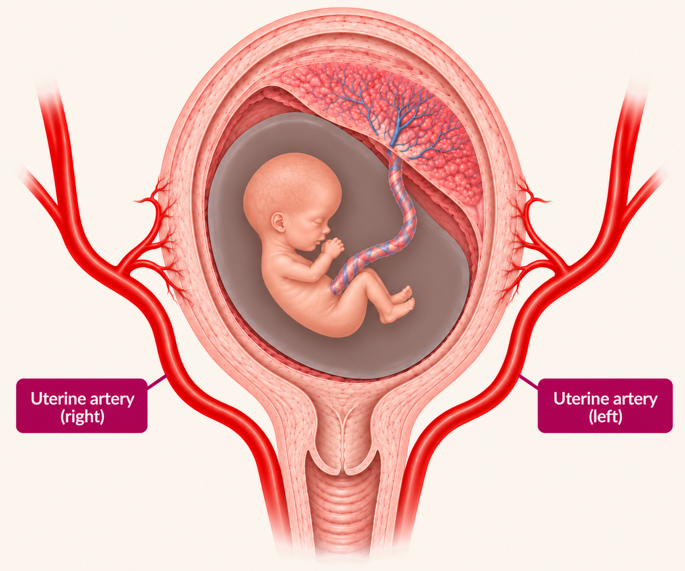
Doppler is an ultrasound-based technique that examines how blood flows through blood vessels. Uterine artery Doppler uses it to look at the flow in the uterine arteries, the blood vessels that supply blood to the uterus and the placenta, and measures the resistance to blood flow in them.
It is a safe, non-invasive examination that can be performed during the 12-week scan and again, where appropriate, at the 20-week scan.
© The Pregnancy Clinic
How does the blood supply to the placenta develop?
Before pregnancy and in early pregnancy, the small blood vessels in the lining of the uterus, called the spiral arteries, are thick-walled and offer resistance to the flow of blood. During the first and early second trimester, these vessels are remodelled and transformed into wide, thin-walled vessels that offer much less resistance, so much more blood can flow to the placenta and the baby.
In some pregnancies, this transformation does not take place fully. High-resistance blood vessels then remain, limiting blood flow to the placenta and, eventually, to the baby. This is associated with pre-eclampsia and fetal growth restriction.
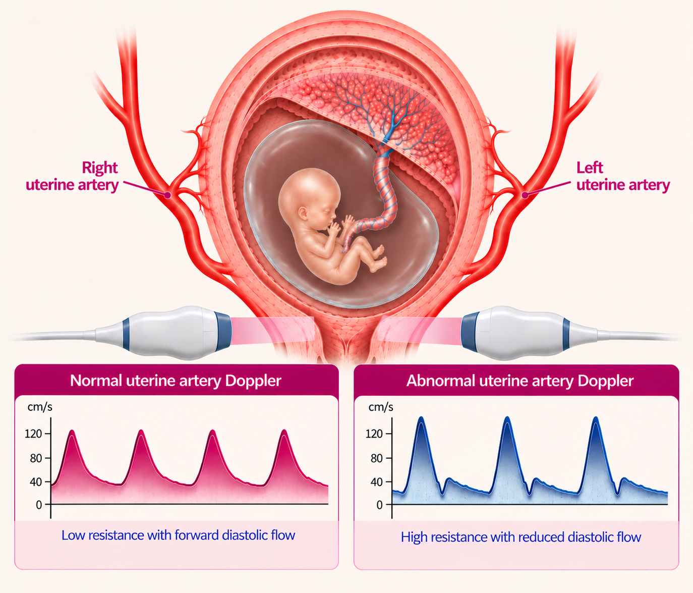
Uterine artery Doppler examines this resistance to blood flow, identifying pregnancies with normal resistance and those with high resistance.
How is it performed?
Uterine artery Doppler is carried out during your 12-week scan. Using colour Doppler, we identify the uterine artery on each side of the uterus and record the pattern of blood flow, which appears as a waveform with each heartbeat. Measurements from the right and left arteries are averaged.
- It is non-invasive and painless, with no needles.
- It does not involve any radiation.
- It is performed by trained specialists using recommended safety settings.
© The Pregnancy Clinic
What does it measure?
The main measurement is the uterine artery pulsatility index (PI), which reflects the resistance to blood flow further downstream. A higher PI indicates greater resistance, and a lower PI lower resistance.
Because the PI normally falls as pregnancy progresses, it is interpreted in relation to the stage of pregnancy and your individual characteristics. A raised PI does not mean that the placenta is failing; it indicates higher resistance, which is associated with a greater chance of certain complications.
Why is uterine artery Doppler performed at 12 weeks?
Some women who later develop pre-eclampsia already have higher resistance in the uterine arteries at 11–13 weeks. On its own, however, uterine artery Doppler is not accurate enough to identify everyone at risk, so it is combined with other measurements in the Fetal Medicine Foundation (FMF) first-trimester screening model:
- Maternal characteristics and history, such as previous pregnancies and medical conditions.
- Mean arterial pressure (MAP), a standardised measurement of your blood pressure.
- Uterine artery PI, measured during the scan.
- A blood test for a marker of placental function, such as placental growth factor (PlGF) or PAPP-A.
Together, these give an individualised risk of pre-eclampsia, particularly preterm pre-eclampsia requiring delivery before 37 weeks. Combined screening identifies around 75% of women who develop preterm pre-eclampsia, at a screen-positive rate of about 10%.
For women identified as high risk, the ASPRE trial showed that aspirin 150 mg at night, started between 11 and 14 weeks and continued until 36 weeks, substantially reduced preterm pre-eclampsia. The main purpose of uterine artery Doppler at 12 weeks is therefore early risk assessment and prevention, rather than diagnosing an existing placental problem.
For further information, read: Pre-eclampsia Screening at 12 Weeks: Can We Predict and Prevent It?
What does a raised result mean?
A raised result means that resistance to blood flow is higher than expected for this stage of pregnancy. It is associated with a higher chance of pre-eclampsia and fetal growth restriction, but many women with a raised result go on to have uncomplicated pregnancies and healthy babies. It should be regarded as a risk marker, not a diagnosis.
Importantly, we do not act on uterine artery Doppler in isolation. We review it as part of your combined pre-eclampsia screening result, together with your history, blood pressure and blood test. If your overall risk is increased, we recommend aspirin 150 mg at night, ideally started at 12–13 weeks and before 16 weeks, and plan appropriate follow-up. A raised PI on its own does not automatically mean that aspirin is needed.
What if my result is normal?
A normal result is reassuring, as it suggests that resistance to blood flow is within the expected range. However, it does not guarantee that pre-eclampsia or fetal growth restriction will not develop, so routine antenatal care, including blood pressure checks, remains important. The most accurate picture comes from combining the Doppler result with your other screening markers and later scan findings.
What is uterine artery notching?
The Doppler waveform sometimes shows a small dip in early diastole, the phase when the heart relaxes, known as an early diastolic notch. Notching is a poor and inaccurate assessment of resistance: many pregnancies with notching do not have increased resistance, and many with increased resistance do not show notching, so it is not reliable. Resistance is much better reflected by the measured pulsatility index.
Moreover, notching is a very common finding at 12 weeks, because the placental circulation is still developing, and on its own it is not a cause for concern.
Is it the same as umbilical artery Doppler?
No. Uterine artery Doppler examines blood flow in the mother's arteries supplying the uterus and placenta. Umbilical artery Doppler examines blood flow in the baby's umbilical cord and is mainly used later in pregnancy when there are concerns about the baby's growth. The two provide complementary information but are not interchangeable.
What about uterine artery Doppler at 20 weeks?
Uterine artery Doppler may also be performed at the 20–22-week anatomy scan, where the purpose is different. By then, resistance should normally have fallen, and persistently raised resistance helps identify pregnancies that may benefit from closer monitoring of the baby's growth later in pregnancy.
| 12-week scan | 20–22-week scan | |
|---|---|---|
| Main purpose | Early screening for pre-eclampsia | Assessing placental blood flow later in pregnancy |
| How it is interpreted | Combined with history, blood pressure and a blood test | Against reference ranges for the stage of pregnancy |
| How it helps | Identifies women who may benefit from aspirin | Guides the need for growth scans |
At 12 weeks, the emphasis is on predicting and preventing complications; at 20 weeks, it is on planning appropriate monitoring. We cover the 20-week examination in detail in a separate guide.
Our approach at The Pregnancy Clinic
At The Pregnancy Clinic, uterine artery Doppler is integrated into our consultant-led 12-week assessment, alongside a detailed examination of your baby. Our assessment includes:
- First-trimester pre-eclampsia screening, combining your history, blood pressure, uterine artery Doppler and a blood test.
- Interpretation in the context of any previous pregnancy complications or medical conditions.
- Clear advice about aspirin where your risk is increased.
- A personalised plan for follow-up, including uterine artery Doppler at 20 weeks and growth scans where indicated, shared with your NHS maternity team.
Uterine artery Doppler is most useful when it is interpreted alongside all the other findings from your pregnancy assessment.
Related pregnancy guides
Frequently asked questions
What is uterine artery Doppler?
It is a safe ultrasound measurement of the resistance to blood flow in the arteries supplying the uterus and placenta, used to assess how the placental blood supply is developing.
Why is uterine artery Doppler done at 12 weeks?
At 11–13 weeks it is combined with your history, blood pressure and a blood test to calculate your risk of pre-eclampsia, so that aspirin can be offered early to women at high risk.
What does a high uterine artery PI mean?
It means resistance to blood flow is higher than expected. It is a risk marker for pre-eclampsia and growth problems, not a diagnosis, and many women with a raised PI have healthy pregnancies.
Is uterine artery notching at 12 weeks normal?
Yes. Notching is very common at 12 weeks and is not a reliable measure of resistance; the measured pulsatility index is far more accurate, so notching on its own is not a concern.
Is uterine artery Doppler safe for my baby?
Yes. It is a non-invasive ultrasound measurement performed with recommended safety settings, and it is performed during your 12-week scan.
Specialist pregnancy ultrasound at The Pregnancy Clinic
At The Pregnancy Clinic, we provide consultant-led ultrasound assessments, including uterine artery Doppler and individualised assessment of placental health, as part of our first-trimester screening, detailed anatomy scans and specialist pregnancy monitoring.
Our approach focuses on clear explanations, careful assessment and personalised care, coordinated with your NHS maternity team.
Book a specialist first-trimester assessment →This guide provides general information and does not replace advice from your maternity care team. Aspirin should only be taken following appropriate clinical assessment. The Pregnancy Clinic provides planned appointments only; for urgent concerns, please contact your maternity unit, NHS 111 or your GP.
Clinical references
- Akolekar R, Syngelaki A, Poon L, Wright D, Nicolaides KH. Competing risks model in early screening for preeclampsia by biophysical and biochemical markers. Fetal Diagn Ther. 2013;33(1):8–15.
- O'Gorman N, Wright D, Syngelaki A, Akolekar R, Wright A, Poon LC, Nicolaides KH. Competing risks model in screening for preeclampsia by maternal factors and biomarkers at 11–13 weeks gestation. Am J Obstet Gynecol. 2016;214(1):103.e1–103.e12.
- Rolnik DL, Wright D, Poon LC, O'Gorman N, Syngelaki A, de Paco Matallana C, Akolekar R, et al. Aspirin versus placebo in pregnancies at high risk for preterm preeclampsia. N Engl J Med. 2017;377(7):613–622.
- Tan MY, Syngelaki A, Poon LC, Rolnik DL, O'Gorman N, Delgado JL, Akolekar R, et al. Screening for pre-eclampsia by maternal factors and biomarkers at 11–13 weeks' gestation. Ultrasound Obstet Gynecol. 2018;52(2):186–195.
- Poon LC, Wright D, Thornton S, Akolekar R, Brocklehurst P, Nicolaides KH. Mini-combined test compared with NICE guidelines for early risk-assessment for pre-eclampsia: the SPREE diagnostic accuracy study. Efficacy Mech Eval. 2020;7(8). NIHR Journals Library.
- Plasencia W, Maiz N, Bonino S, Kaihura C, Nicolaides KH. Uterine artery Doppler at 11+0 to 13+6 weeks in the prediction of pre-eclampsia. Ultrasound Obstet Gynecol. 2007;30(5):742–749.
- Plasencia W, Maiz N, Poon L, Yu C, Nicolaides KH. Uterine artery Doppler at 11+0 to 13+6 weeks and 21+0 to 24+6 weeks in the prediction of pre-eclampsia. Ultrasound Obstet Gynecol. 2008;32(2):138–146.
- National Institute for Health and Care Excellence. Hypertension in pregnancy: diagnosis and management (NG133).
- NHS England. Saving babies' lives version three: a care bundle for reducing perinatal mortality.
- Fetal Medicine Foundation. Uterine artery Doppler and first-trimester pre-eclampsia screening.