
Discovering that you are expecting twins is an exciting moment, although it can also bring additional questions about your pregnancy.
Most twin pregnancies are identified during an early pregnancy scan, usually between 6 and 8 weeks. However, the 12-week scan is particularly important because it establishes the type of twin pregnancy and helps determine the care you will need throughout pregnancy.
Key points
- The 12-week scan establishes whether your babies share a placenta, known as chorionicity.
- Chorionicity determines how often you will need scans and helps identify pregnancies that need closer monitoring.
- Screening for Down's, Edwards' and Patau's syndromes is available in twin pregnancies.
- Twins sharing a placenta (monochorionic twins) need scans every 2 weeks from 16 weeks.
- Although twin pregnancies have higher risks than singleton pregnancies, most result in healthy babies.
What can the 12-week scan tell you about your twins?
At The Pregnancy Clinic, our detailed twin pregnancy scan at 11 to 14 weeks assesses whether your babies share a placenta or amniotic sac, their size and early anatomical development, their nuchal translucency measurements and the screening options available for chromosomal conditions.
The findings help us identify pregnancies that may need closer monitoring or specialist fetal medicine care.
What are the different types of twins?
Twin pregnancies are classified according to the number of placentas and amniotic sacs.
- Chorionicity describes whether the babies have separate placentas or share one placenta.
- Amnionicity describes whether each baby has a separate amniotic sac or whether both babies share the same sac.
There are three main types of twin pregnancy.
| Type | Placenta | Amniotic sacs |
|---|---|---|
| Dichorionic diamniotic (DCDA) | Two separate placentas | Two separate sacs |
| Monochorionic diamniotic (MCDA) | One shared placenta | Two separate sacs |
| Monochorionic monoamniotic (MCMA) | One shared placenta | One shared sac |
DCDA twins are the most common type. MCDA twins need closer monitoring because they share a placental circulation, while MCMA twins are rare and need particularly specialised care.
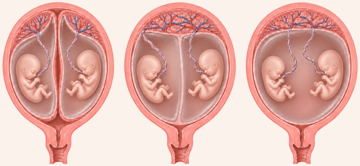
diamnioticMonochorionic
diamnioticMonochorionic
monoamniotic
© The Pregnancy Clinic
How are twin pregnancies formed?
In a typical menstrual cycle, one egg is released from the ovary. When this egg is fertilised by a sperm, a single embryo develops. The embryo implants in the womb and usually forms a pregnancy with one baby, one placenta and one amniotic sac, known as a singleton pregnancy.
Twin pregnancies can develop in two different ways.
Two eggs fertilised by two sperm: non-identical twins
Occasionally, two eggs are released during the same menstrual cycle. If both eggs are fertilised by separate sperm, two embryos develop and implant in the womb. Each embryo forms its own placenta and amniotic sac, resulting in dichorionic diamniotic (DCDA) twins.
Because the embryos come from different eggs and sperm, they have different genetic make-ups, just like other brothers and sisters. These are called dizygotic twins, commonly known as non-identical or fraternal twins. They may be the same sex or different sexes.
One fertilised egg divides into two embryos: identical twins
Occasionally, a single egg fertilised by one sperm divides to form two embryos. Because both embryos come from the same fertilised egg, they generally have the same genetic make-up. These are called monozygotic twins, commonly known as identical twins.
The number of placentas and amniotic sacs depends largely on when the embryo divides.
| Approximate timing of division | Type of twins formed | Placenta and sacs |
|---|---|---|
| Within the first 3 days | Dichorionic diamniotic (DCDA) | Separate placentas and sacs |
| Around days 4 to 8 | Monochorionic diamniotic (MCDA) | Shared placenta, separate sacs |
| After about day 8 | Monochorionic monoamniotic (MCMA) | Shared placenta and shared sac |
This is the traditional explanation of how identical twins develop. The timings are approximate, and the precise biological mechanisms are more complex. Identical twins can therefore have separate placentas, share a placenta but have separate amniotic sacs, or share both the placenta and the amniotic sac.
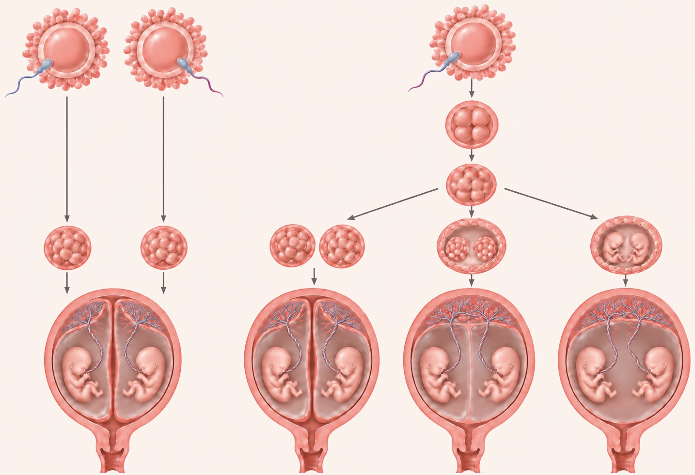
diamnioticDichorionic
diamnioticMonochorionic
diamnioticMonochorionic
monoamniotic
© The Pregnancy Clinic
What is the difference between zygosity and chorionicity?
Zygosity describes whether the twins developed from one fertilised egg or two. Monozygotic twins come from one fertilised egg and are generally genetically identical, while dizygotic twins come from two separate fertilised eggs and are genetically different.
Chorionicity describes whether the twins share a placenta. Ultrasound can determine chorionicity reliably in the first trimester, but it cannot always establish zygosity.
- Monochorionic twins are almost always monozygotic and therefore identical.
- Dichorionic twins of different sexes are dizygotic and therefore non-identical in ordinary circumstances.
- Dichorionic twins of the same sex may be either monozygotic or dizygotic.
If twins have separate placentas and are the same sex, ultrasound alone cannot reliably determine whether they are identical. Genetic testing can establish zygosity when needed.
For pregnancy care, chorionicity is generally more important than zygosity, because sharing a placenta is associated with additional pregnancy risks.
How is chorionicity determined on ultrasound?
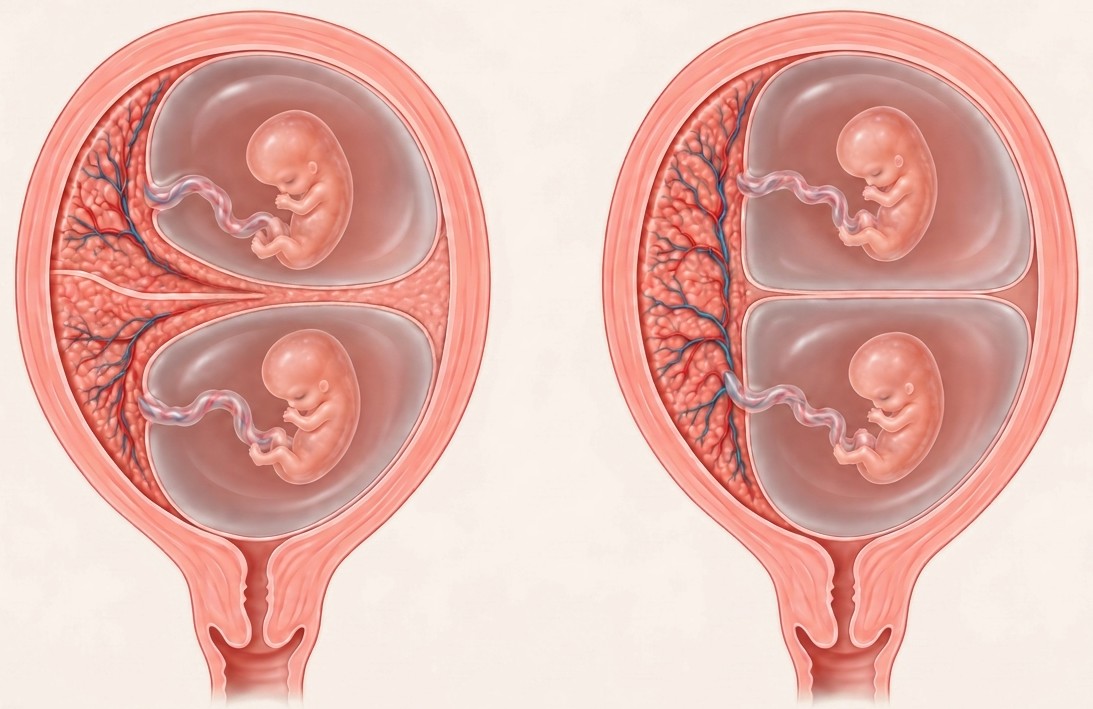
The 11 to 14-week scan is the best opportunity to determine chorionicity accurately. During the scan, we assess the number and appearance of the placentas, the membrane separating the babies and the way this membrane joins the placenta. Two important ultrasound signs are used.
- The lambda sign, also called the twin-peak sign, is a small triangular projection of placental tissue where the membrane separating the twins joins the placenta. Its presence indicates a dichorionic twin pregnancy.
- The T-sign occurs when a thin membrane separating the twins meets a shared placenta at approximately a right angle. This indicates a monochorionic diamniotic twin pregnancy.
In monochorionic monoamniotic twins, there is no membrane separating the babies. The number of visible placentas alone is not always reliable, because two separate placentas may lie next to each other and appear as a single placental mass. At The Pregnancy Clinic, we carefully examine these features and document chorionicity during the first-trimester scan.
© The Pregnancy Clinic
How is the due date calculated in twins?
At the 12-week scan, we measure the crown–rump length (CRL) of both babies. In spontaneously conceived twin pregnancies, gestational age is generally calculated using the larger baby's crown–rump length. This avoids assigning an incorrect gestational age if one baby is smaller because of an early growth difference.
In IVF pregnancies, the known date of fertilisation or embryo transfer is used to establish the gestational age. Both babies are measured individually, and any significant difference in size is documented.
For further information, read: How Accurate Is My Due Date? Pregnancy Dating Explained
Can twins be screened for Down's syndrome?
Yes. Screening for Down's syndrome, Edwards' syndrome and Patau's syndrome is available in twin pregnancies.
- Combined first-trimester screening uses the nuchal translucency measurement of each baby, maternal blood tests and other relevant information to estimate the chance of chromosomal conditions.
- Non-invasive prenatal testing (NIPT) analyses cell-free DNA in the mother's blood and provides highly accurate screening for Down's syndrome. NIPT can be performed in twin pregnancies, although its performance and the information available depend on the type of twin pregnancy and the laboratory.
If one twin has stopped developing, sometimes called a vanishing twin, NIPT results may be more difficult to interpret. When screening shows a higher chance of a chromosomal condition, or an ultrasound abnormality is found, diagnostic testing with CVS or amniocentesis may be discussed.
For further information, read: NIPT vs Combined Screening: Which Test Should I Choose?
What are the complications of twin pregnancies?
Most twin pregnancies result in healthy babies. However, compared with pregnancies with one baby, twins have a higher chance of certain complications, including miscarriage, stillbirth, fetal abnormalities, preterm birth, fetal growth restriction and pre-eclampsia. The chance of complications also differs between dichorionic and monochorionic twins.
How do pregnancy risks compare?
| Pregnancy complicationComplication | Singleton pregnancySingleton | Dichorionic twinsDC twins | Monochorionic twinsMC twins |
|---|---|---|---|
| Miscarriage (11–23 weeks) | 1% | 2% | 8% |
| Fetal death (from 24 weeks) | 0.3% | 2% | 4% |
| Fetal growth restriction | 10% | 20% | 30% |
| Preterm birth (before 32 weeks) | 0.8% | 5% | 10% |
| Major defects | 0.5% | 1% | 4% |
These figures are approximate. Miscarriage figures refer to pregnancy loss after a reassuring scan at 11 to 14 weeks, rather than the chance of miscarriage from conception. DC: dichorionic; MC: monochorionic.
Overall, about 60% of twin pregnancies result in birth before 37 weeks, although this includes both spontaneous and planned early deliveries. The chance of individual complications depends on your health, pregnancy history, chorionicity and the findings of ultrasound monitoring.
Why are monochorionic twins higher risk?
Monochorionic twins share a single placenta. Within this placenta, blood vessels from the two babies are usually connected, allowing blood to pass between their circulations. In most pregnancies, this circulation remains balanced enough for both babies to develop normally. However, the shared placenta can occasionally lead to complications that do not occur in the same way in dichorionic twins.
- Twin-to-twin transfusion syndrome (TTTS): blood flow between the twins becomes unbalanced. One baby may have a reduced blood volume and very little amniotic fluid, while the other has an increased blood volume and too much amniotic fluid. TTTS needs specialist assessment and, in some cases, treatment during pregnancy.
- Selective fetal growth restriction: the shared placenta may not be divided equally. If one baby receives a smaller or less effective share, that baby may grow more slowly than the other.
- Twin anaemia–polycythaemia sequence (TAPS): small blood vessel connections allow a slow transfer of blood between the twins. One baby may become anaemic, while the other develops an increased concentration of red blood cells.
- Greater consequences if one twin becomes unwell: because the twins share blood vessel connections, a serious complication affecting one baby may also affect the other.
This is why monochorionic pregnancies need more frequent ultrasound monitoring. The increased risks are related mainly to the shared placenta and its circulation, rather than simply to the twins being genetically identical.
Will I need more scans because I am expecting twins?
Yes. All twin pregnancies need additional ultrasound monitoring, and the frequency depends mainly on chorionicity.
| Type of pregnancy | Usual ultrasound monitoring |
|---|---|
| Dichorionic diamniotic twins | Growth scans about every 4 weeks from 24 weeks |
| Monochorionic diamniotic twins | Scans every 2 weeks from 16 weeks |
| Monochorionic monoamniotic twins | Individualised specialist monitoring |
These scans monitor the babies' growth, the amniotic fluid and other findings that may indicate developing complications.
Twin pregnancies also have a higher chance of preterm birth. We recommend cervical length assessment from 16 weeks if you have other risk factors for preterm birth in addition to twins, such as a previous preterm birth or cervical surgery. If you have no additional risk factors, we offer cervical length assessment at 20 to 22 weeks. This helps identify women who may benefit from preventive treatment. We also assess your risk factors for pre-eclampsia and discuss whether low-dose aspirin is appropriate.
Related pregnancy guides
Frequently asked questions
When can you tell if twins share a placenta?
Chorionicity is best determined at the 11 to 14-week scan, using the lambda sign and the T-sign where the membrane between the twins joins the placenta.
Are twins who share a placenta always identical?
Monochorionic twins, who share a placenta, are almost always identical. Twins with separate placentas may be identical or non-identical.
Can I have NIPT with twins?
Yes. NIPT can be performed in twin pregnancies, although its performance depends on the type of twin pregnancy and the laboratory, and results can be harder to interpret if one twin has stopped developing.
How is the due date worked out for twins?
In spontaneously conceived twins, the due date is generally calculated from the larger baby's crown–rump length at the 12-week scan. In IVF pregnancies, the known dates of conception are used.
How many scans will I need with twins?
Dichorionic twins usually have growth scans about every 4 weeks from 24 weeks. Monochorionic twins need scans every 2 weeks from 16 weeks.
Are twin pregnancies high risk?
Twin pregnancies have higher risks than singleton pregnancies, particularly when the twins share a placenta, but most result in healthy babies.
Twin pregnancy scans at The Pregnancy Clinic
Our consultant-led first-trimester twin scan provides a detailed assessment of both babies, including:
- Accurate determination of chorionicity and amnionicity.
- Assessment of the size and early anatomy of both babies.
- Nuchal translucency measurements and discussion of chromosomal screening.
- Assessment of significant differences between the twins.
- Review of pregnancy risks and recommendations for subsequent monitoring.
We take time to explain the type of twin pregnancy, what the findings mean and how your care should be planned throughout pregnancy.
Book your 12-week twin pregnancy scan →An additional twin pregnancy charge of £100 applies per scan. This guide provides general information and does not replace personalised medical advice.
Clinical references
- Sepulveda W, Sebire NJ, Hughes K, Odibo A, Nicolaides KH. The lambda sign at 10–14 weeks of gestation as a predictor of chorionicity in twin pregnancies. Ultrasound Obstet Gynecol 1996;7:421–3
- Litwinska E, Syngelaki A, Cimpoca B, Frei L, Nicolaides KH. Outcome of twin pregnancy with two live fetuses at 11–13 weeks' gestation. Ultrasound Obstet Gynecol 2020;55:32–8
- Syngelaki A, Cimpoca B, Litwinska E, Akolekar R, Nicolaides KH. Diagnosis of fetal defects in twin pregnancies at routine 11–13-week ultrasound examination. Ultrasound Obstet Gynecol 2020;55:474–81
- Judah H, Gil MM, Syngelaki A, Galeva S, Jani J, Akolekar R, Nicolaides KH. Cell-free DNA testing of maternal blood in screening for trisomies in twin pregnancy: updated cohort study at 10–14 weeks and meta-analysis. Ultrasound Obstet Gynecol 2021;58:178–89
- Galeva S, Konstantinidou L, Gil MM, Akolekar R, Nicolaides KH. Routine first-trimester screening for fetal trisomies in twin pregnancy: cell-free DNA test contingent on results from combined test. Ultrasound Obstet Gynecol 2019;53:208–13
- Wright A, Wright D, Chaveeva P, Molina FS, Akolekar R, Syngelaki A, Petersen OB, Kristensen SE, Nicolaides KH. Fetal Medicine Foundation charts for fetal growth in twins. Ultrasound Obstet Gynecol 2024;63:181–8
- NICE — Twin and triplet pregnancy (NG137)
- RCOG — Monochorionic twin pregnancy, management (Green-top Guideline No. 51)
- RCOG — Multiple pregnancy: having more than one baby (patient information)
- NHS — Screening for Down's syndrome, Edwards' syndrome and Patau's syndrome