Nuchal translucency (NT) is one of the measurements obtained during the first-trimester pregnancy scan. It refers to a small, translucent area containing fluid beneath the skin at the back of the baby's neck. Every baby has some nuchal translucency, and in most pregnancies the amount of fluid is within the expected range for the baby's size.
Occasionally, the measurement is greater than expected. An increased nuchal translucency does not necessarily mean that your baby has a problem, but it is an important finding that warrants careful specialist assessment and consideration of further investigations.
Key points
- NT is measured between 11 weeks and 13 weeks 6 days, when the baby's crown–rump length is about 45–84 mm.
- There is no single normal value: NT increases with the baby's size and is interpreted against reference ranges.
- An increased NT may be linked with chromosomal or genetic conditions or heart defects, but can also be a normal variation.
- Many pregnancies with increased NT have a favourable outcome when further scans and genetic tests are reassuring.
What is nuchal translucency?
Nuchal translucency is a translucent, fluid-filled area beneath the skin at the back of the baby's neck that can be seen on ultrasound in early pregnancy. It is a normal part of fetal development, and the amount of fluid normally increases as the baby grows.
When the amount of fluid is greater than expected for the baby's size, this is described as an increased nuchal translucency. It can be associated with chromosomal or genetic conditions and with structural abnormalities, particularly of the heart, but it can also represent a normal variation with no underlying abnormality.

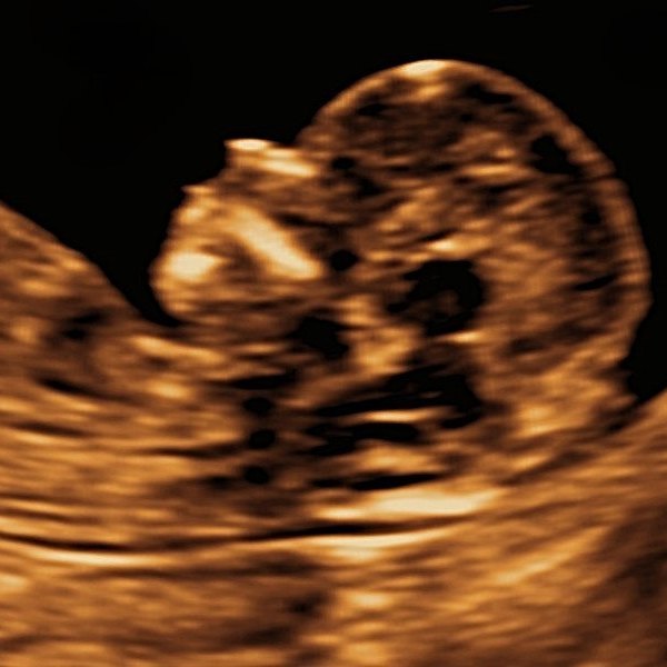
Fetal nuchal translucency at 12 weeks
© The Pregnancy Clinic
When and how is NT measured?
NT is measured between 11 weeks and 13 weeks plus 6 days, when the baby's crown–rump length (CRL) measures approximately 45–84 mm.
The measurement requires a specific image showing the baby side-on, with the head and neck in a neutral position. The clinician measures the widest part of the fluid-filled space behind the neck, taking care to distinguish the baby's skin from the surrounding amniotic membrane. It is usually obtained by abdominal ultrasound; occasionally a transvaginal scan gives clearer images.
Accurate measurement matters, because differences of less than a millimetre can change the interpretation of the result.
What is a normal NT measurement?
There is no single NT measurement that is normal for every baby. Because NT increases as the baby grows, it must be interpreted in relation to the crown–rump length rather than gestational age alone.
Most babies have NT measurements below approximately 2.5–3.0 mm during the screening window, but these figures are not fixed thresholds:
- A measurement above the 95th centile for the baby's CRL is generally described as increased.
- An NT of 3.5 mm or more is commonly used as a threshold for more detailed assessment, regardless of the baby's size.
- A measurement below 3.5 mm may still be above the expected centile for a smaller baby.
What does an increased NT mean?
There are several possible explanations. In some pregnancies, the extra fluid is a temporary variation in normal development that resolves as pregnancy progresses. In others, it may be associated with chromosomal conditions, genetic syndromes or structural abnormalities, particularly congenital heart defects. The likelihood of an underlying condition increases as the NT measurement becomes larger.
An increased NT is not a diagnosis. It indicates the need for further specialist assessment, which may include a detailed examination of the baby's anatomy and heart, additional genetic screening or diagnostic testing.
Chromosomal conditions
An increased NT is associated with a higher chance of trisomy 21 (Down's syndrome), trisomy 18 (Edwards' syndrome), trisomy 13 (Patau's syndrome), and Turner syndrome and other sex chromosome abnormalities. However, most babies with Down's syndrome do not have a markedly enlarged NT, and many babies with an increased NT have normal chromosomes. For this reason, NT is combined with your age and the blood markers PAPP-A and free β-hCG in combined screening. NIPT offers more accurate screening for the common trisomies but does not identify every genetic condition.
Heart abnormalities
Congenital heart disease is one of the important associations of increased NT, even when the chromosomes are normal, and the likelihood of a major heart defect increases with the degree of NT enlargement. With specialist expertise, some major heart abnormalities can be identified at 12–13 weeks, and an early cardiac assessment and later fetal echocardiogram may be recommended. A normal chromosome result does not remove the need for detailed cardiac assessment when the NT is significantly increased.
Other genetic conditions
Occasionally, increased NT is associated with single-gene disorders, particularly conditions affecting the heart, lymphatic system or other organs. One recognised group is the RASopathies, which include Noonan syndrome. The need for additional genetic testing depends on the degree of NT enlargement, whether other abnormalities are present and the results of initial investigations.
What happens if my baby's NT is increased?
The next steps depend on the NT measurement, the baby's crown–rump length, any other ultrasound findings and any screening results already available. A specialist assessment usually includes a detailed review of the baby's anatomy, particularly the heart, brain and abdominal wall. Further investigations may be discussed:
- NIPT provides highly accurate screening for Down's, Edwards' and Patau's syndromes, but does not exclude all chromosomal or genetic conditions and cannot replace diagnostic testing when there is a significant ultrasound finding.
- Chorionic villus sampling (CVS), usually from 11 weeks, analyses a small sample of placental tissue.
- Amniocentesis, usually from 15 weeks, analyses a sample of amniotic fluid.
Diagnostic testing may include chromosome analysis or chromosomal microarray and, in selected cases, further molecular genetic testing. When NT is substantially increased, diagnostic testing can provide information that NIPT alone cannot. The benefits, limitations and risks of each option should be discussed before a decision is made.
What if the chromosome results are normal?
A normal chromosome result is reassuring. Depending on the degree of NT enlargement, a detailed scan in the early second trimester and a specialist fetal heart examination may be recommended, and the 20-week anatomy scan remains important. If the NT was markedly increased or other findings are present, further genetic assessment may still be considered.
When the NT returns to the expected range, the baby's anatomy and heart are normal and the relevant genetic investigations are reassuring, the outlook is often favourable.
Can increased nuchal translucency disappear?
Yes. In many pregnancies the fluid becomes less prominent as pregnancy progresses, which is generally reassuring. However, its disappearance does not retrospectively exclude a chromosomal, genetic or structural condition, so if the original measurement was significantly increased, the recommended investigations and follow-up should still be considered.
Does a normal NT mean my baby is healthy?
A normal NT is reassuring but cannot guarantee that a baby does not have a chromosomal, genetic or structural abnormality. Some babies with Down's syndrome have NT measurements within the normal range, and many heart defects are not associated with increased NT.
NT is therefore only one part of first-trimester assessment. A detailed first-trimester scan, appropriate chromosomal screening and the fetal anatomy scan at 20 weeks provide complementary information about your baby's development.
Related pregnancy guides
Frequently asked questions
What is a normal nuchal translucency measurement?
There is no single normal value, because NT increases with the baby's size. Most babies measure below about 2.5–3.0 mm; a measurement above the 95th centile for the baby's crown–rump length is described as increased.
What NT measurement is considered high?
A measurement above the 95th centile for the baby's size is increased, and an NT of 3.5 mm or more is commonly used as a threshold for more detailed specialist assessment.
Does increased NT mean my baby has Down's syndrome?
No. Increased NT raises the chance of chromosomal conditions, but many babies with increased NT have normal chromosomes. It indicates the need for further assessment, not a diagnosis.
Can increased NT indicate a heart problem?
Yes. Congenital heart defects are an important association, even when the chromosomes are normal, so a detailed heart assessment may be recommended.
Is cystic hygroma the same as increased NT?
A cystic hygroma is a type of increased nuchal translucency. It is a lymphatic abnormality that usually appears as a more extensive collection of fluid around the baby's neck, often with internal divisions (septations). It may be associated with chromosomal abnormalities, genetic syndromes or abnormal lymphatic development, and warrants specialist fetal medicine assessment.
Can increased NT go away?
Yes, the fluid often becomes less prominent later in pregnancy. This is reassuring, but the recommended follow-up should still be considered if the original measurement was significantly increased.
Does a normal NT mean my baby is healthy?
A normal NT is reassuring but cannot exclude every condition. A detailed scan, chromosomal screening and the 20-week fetal anatomy scan give complementary information.
First-trimester pregnancy care at The Pregnancy Clinic
At The Pregnancy Clinic, we provide consultant-led first-trimester ultrasound examinations, including detailed assessment of nuchal translucency and early fetal anatomy. Where an increased NT measurement is identified, we provide specialist assessment of the developing baby, explain the significance of the findings and discuss the available screening and diagnostic investigations.
Our approach is to provide clear, evidence-based information and individualised counselling, allowing parents sufficient time to understand their results and consider the next steps.
Book a first-trimester consultation →An increased NT measurement requires interpretation alongside the baby's size, ultrasound findings and relevant screening results. Further investigations should be individualised following specialist assessment. The Pregnancy Clinic provides planned appointments only; for urgent concerns, please contact your local NHS maternity unit, Early Pregnancy Unit, NHS 111 or your GP.
Clinical references
- Minnella GP, Crupano FM, Syngelaki A, Zidere V, Akolekar R, Nicolaides KH. Diagnosis of major heart defects by routine first-trimester ultrasound examination: association with high nuchal translucency, tricuspid regurgitation and abnormal flow in the ductus venosus. Ultrasound Obstet Gynecol. 2020;55(5):637–644.
- Nicolaides KH. Nuchal translucency and other first-trimester sonographic markers of chromosomal abnormalities. Am J Obstet Gynecol. 2004;191(1):45–67.
- Wright D, Kagan KO, Molina FS, Gazzoni A, Nicolaides KH. A mixture model of nuchal translucency thickness in screening for chromosomal defects. Ultrasound Obstet Gynecol. 2008;31(4):376–383.
- Souka AP, von Kaisenberg CS, Hyett JA, Sonek JD, Nicolaides KH. Increased nuchal translucency with normal karyotype. Am J Obstet Gynecol. 2005;192(4):1005–1021.
- Hyett J, Perdu M, Sharland G, Snijders R, Nicolaides KH. Using fetal nuchal translucency to screen for major congenital cardiac defects at 10–14 weeks of gestation: population based cohort study. BMJ. 1999;318(7176):81–85.
- Bilardo CM, Chaoui R, Hyett JA, Kagan KO, Karim JN, Papageorghiou AT, et al. ISUOG Practice Guidelines (updated): performance of 11–14-week ultrasound scan. Ultrasound Obstet Gynecol. 2023;61(1):127–143.
- NHS. Screening for Down's syndrome, Edwards' syndrome and Patau's syndrome.
- National Institute for Health and Care Excellence. Antenatal care (NG201). 2021.