During the first trimester, expectant parents are usually offered screening for three common chromosomal conditions: Down's syndrome (trisomy 21), Edwards' syndrome (trisomy 18) and Patau's syndrome (trisomy 13). Two principal approaches are available: combined first-trimester screening and non-invasive prenatal testing (NIPT).
Both estimate the chance of the baby having these conditions, but they use different methods and have different levels of accuracy. Importantly, neither test replaces a detailed ultrasound examination of the baby's developing anatomy and heart at 12–13 weeks.
Key points
- Standard NHS combined screening detects around 85–87% of pregnancies affected by Down's syndrome.
- Enhanced specialist screening with additional ultrasound markers can raise detection to around 95–98%.
- NIPT detects around 99–99.7%, with a false-positive rate of only about 0.04–0.1%.
- Both are screening tests: a higher-chance result needs counselling and the offer of diagnostic testing.
What is combined first-trimester screening?
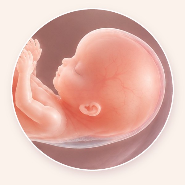
Combined screening is the standard NHS approach. It is performed between 11 weeks and 13 weeks plus 6 days, when the baby's crown–rump length measures approximately 45–84 mm, and combines four factors:
- Maternal age, as the chance of certain chromosomal conditions increases with age.
- Nuchal translucency (NT), the fluid beneath the skin at the back of the baby's neck.
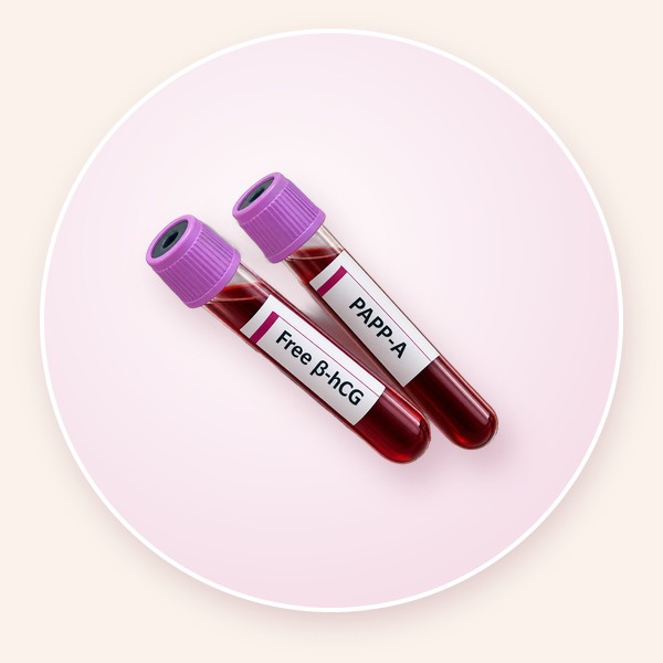
- PAPP-A (pregnancy-associated plasma protein-A), measured in the mother's blood.
- Free β-hCG (free beta-human chorionic gonadotrophin), a hormone measured in the mother's blood.
These are combined in a validated algorithm to give an individual chance, such as 1 in 100 or 1 in 5,000. Combined screening does not diagnose a chromosomal condition; it identifies pregnancies with a higher or lower chance of an affected baby.
How accurate is it?
For Down's syndrome, the standard NHS programme has a reported detection rate of approximately 85–87%. This means around 13–15% of affected pregnancies receive a lower-chance result (false negatives), while some unaffected pregnancies receive a higher-chance result (false positives). Detection rates for Edwards' and Patau's syndromes differ from those for Down's syndrome.
Can combined screening be made more accurate?
Yes. Detection of Down's syndrome can be improved from approximately 85–87% with standard NHS combined screening to around 95–98% with enhanced first-trimester screening in experienced specialist centres. A comprehensive fetal medicine assessment can add:
- Fetal nasal bone: whether the nasal bone is present and developed.
- Ductus venosus Doppler: blood flow through a small vessel connecting the umbilical circulation to the heart.
- Tricuspid valve Doppler: abnormal blood flow across the tricuspid valve.
Research from the Fetal Medicine Foundation has shown that including these markers in validated algorithms substantially improves detection and reduces false-positive results.
The accuracy of ultrasound-based screening also depends on the training and experience of the clinician. NT must be measured using a standardised technique, and reliable assessment of the additional markers requires specialist expertise. A detailed assessment also includes a systematic examination of the baby's anatomy and heart, which can identify major abnormalities associated with chromosomal or genetic conditions that a basic dating and NT scan may not.
Even enhanced screening cannot detect every affected pregnancy, and a normal scan does not exclude all chromosomal or genetic conditions.
For further information, read: Nuchal Translucency (NT): What Is Normal and What Does It Mean?
What is NIPT?
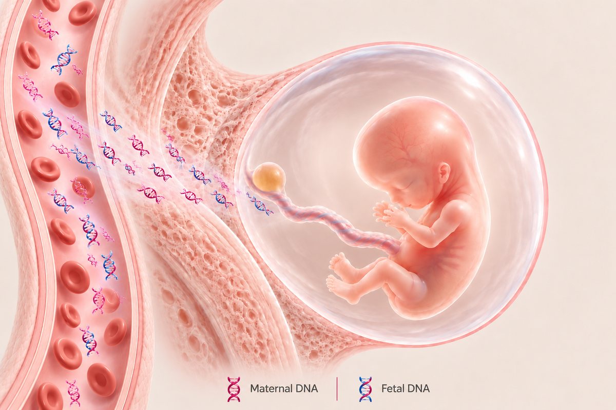
During pregnancy, the placenta releases small fragments of DNA, known as cell-free DNA, into the mother's bloodstream. Because placental DNA usually reflects the baby's chromosomes, analysing these fragments allows laboratories to screen for certain chromosomal conditions with a high degree of accuracy.
© The Pregnancy Clinic
NIPT requires only a blood sample from the mother and can generally be performed from 10 weeks. It does not rely on NT, PAPP-A or free β-hCG, and its principal advantage is substantially higher accuracy, particularly for Down's syndrome.
How accurate is NIPT?
Large studies and systematic reviews report a detection rate of approximately 99–99.7% for trisomy 21, with a false-positive rate of about 0.04–0.1% and a false-negative rate of about 0.3–1%, depending on the test and population.
Even so, NIPT remains a screening test rather than a diagnostic investigation. A lower-chance result is highly reassuring for Down's syndrome but does not exclude every chromosomal, genetic or structural abnormality.
Comparison for Down's syndrome (trisomy 21)
| Screening performance | Standard NHS combined screening | Enhanced specialist combined screening | NIPT |
|---|---|---|---|
| Detection rate | 85–87% | 95–98% | 99–99.7% |
| False-negative rate | 13–15% | 2–5% | 0.3–1% |
| False-positive rate | Approximately 2.3% | Approximately 2.5–3% in published enhanced-marker models | Approximately 0.04–0.1% |
Indicative screening performance for trisomy 21, not a guarantee for an individual pregnancy. Estimates for enhanced screening vary with the markers, algorithms and pathways used. The false-positive rate is the proportion of unaffected pregnancies receiving a higher-chance result.
Higher-chance and lower-chance results
Both tests give an estimate of chance rather than a diagnosis. A lower-chance result means the test has not found evidence of an increased chance; it is reassuring, particularly after NIPT, but does not completely exclude the condition. A higher-chance result means the test has found findings associated with an increased probability; it does not mean the baby definitely has the condition.
After a higher-chance combined result
In the NHS screening programme in England, a combined result of 1 in 150 or higher is classified as higher chance. Options include no further testing, NIPT as an additional screening test, or diagnostic testing with chorionic villus sampling (CVS) or amniocentesis. Where there are significant structural abnormalities or a markedly increased NT, direct diagnostic testing may be more appropriate than NIPT alone.
After a higher-chance NIPT result
NIPT analyses predominantly placental DNA. Occasionally the placenta's chromosomes differ from the baby's, known as confined placental mosaicism, which is one reason false-positive results occur. A higher-chance NIPT result should therefore never be treated as a diagnosis without confirmatory testing: CVS, usually from 11 weeks, or amniocentesis, usually from 15 weeks. The most appropriate test depends on the condition suspected, the scan findings, the gestation and the possibility of placental mosaicism.
What NIPT cannot do
Some NIPT tests also screen for sex chromosome abnormalities or selected microdeletions, but performance for these conditions is not comparable with Down's syndrome, and the chance that a higher-chance result is correct may be substantially lower for rare conditions. NIPT cannot exclude every chromosomal abnormality, single-gene disorder or genetic syndrome.
Occasionally, NIPT gives no result, often because of a low fetal fraction (too little placental DNA in the sample). This can relate to early gestation, maternal characteristics, placental factors or certain chromosomal conditions. A no-result should not be regarded as reassuring; options include repeating the test, a detailed scan or diagnostic testing.
Why the 12-week scan still matters if you choose NIPT
NIPT evaluates the chance of selected chromosomal conditions. It does not examine the baby's developing anatomy. A detailed scan at 12–13 weeks by an experienced specialist can identify major structural abnormalities of the brain, spine, face, heart, abdominal wall, bladder and limbs that NIPT would not detect.
Nuchal translucency also remains important: an increased NT can be associated with heart defects, genetic syndromes and other abnormalities not assessed by NIPT, and may indicate the need for further investigation even when NIPT is reassuring. A normal NIPT result cannot exclude a major structural abnormality, just as a normal scan cannot exclude every chromosomal or genetic condition.
For further information, read: What Fetal Abnormalities Can Be Detected at the 12-Week Scan?
Which is better?
For screening specifically for Down's, Edwards' and Patau's syndromes, NIPT generally offers superior accuracy, with higher detection and fewer false positives. Combined screening remains an established NHS approach, and its performance improves substantially with specialist assessment of additional markers.
The right choice depends on what you wish to know and your individual circumstances. For parents seeking the most accurate screening for the common trisomies alongside detailed information about their baby's development, NIPT combined with a specialist first-trimester scan offers the most comprehensive approach.
Related pregnancy guides
Frequently asked questions
Is NIPT more accurate than combined screening?
Yes. NIPT detects around 99–99.7% of pregnancies affected by Down's syndrome with a false-positive rate of about 0.04–0.1%, compared with around 85–87% for standard NHS combined screening.
Can combined screening be improved?
Yes. Enhanced specialist screening, adding the nasal bone, ductus venosus and tricuspid valve Doppler, can raise detection of Down's syndrome to around 95–98% in experienced centres.
When can NIPT be done?
NIPT can generally be performed from 10 weeks of pregnancy, depending on the laboratory and test used.
Is NIPT a diagnostic test?
No. NIPT is a screening test. A higher-chance result should be confirmed with CVS or amniocentesis before any decisions are made.
Do I still need a 12-week scan if I have NIPT?
Yes. NIPT does not examine the baby's anatomy. A detailed 12-week scan can identify major structural abnormalities and an increased nuchal translucency that NIPT would not detect.
What if NIPT gives no result?
A no-result, often due to low fetal fraction, should not be regarded as reassuring. Options include repeating the test, a detailed scan or diagnostic testing, following specialist advice.
First-trimester screening at The Pregnancy Clinic
At The Pregnancy Clinic, we provide consultant-led first-trimester assessments combining specialist ultrasound expertise with personalised advice about prenatal screening and diagnostic investigations. Our approach includes detailed assessment of early fetal anatomy, examination of the developing heart and evaluation of ultrasound markers associated with chromosomal and genetic conditions.
We also offer a range of NIPT options, so that you can choose the right screening approach after discussing the benefits, accuracy and limitations of each test. Where further investigations are indicated, we provide clear explanations and individualised counselling.
Book a first-trimester scan or NIPT consultation →Prenatal screening does not replace diagnostic testing when clinically indicated, and a normal first-trimester assessment does not replace the recommended mid-pregnancy fetal anatomy scan. The Pregnancy Clinic provides planned appointments only; for urgent concerns, please contact your local NHS maternity unit, Early Pregnancy Unit, NHS 111 or your GP.
Clinical references
- Nicolaides KH. Screening for fetal aneuploidies at 11 to 13 weeks. Prenat Diagn. 2011;31(1):7–15.
- Nicolaides KH. Nuchal translucency and other first-trimester sonographic markers of chromosomal abnormalities. Am J Obstet Gynecol. 2004;191(1):45–67.
- Gil MM, Akolekar R, Quezada MS, Bregant B, Nicolaides KH. Analysis of cell-free DNA in maternal blood in screening for aneuploidies: meta-analysis. Fetal Diagn Ther. 2014;35(3):156–173.
- Gil MM, Accurti V, Santacruz B, Plana MN, Nicolaides KH. Analysis of cell-free DNA in maternal blood in screening for aneuploidies: updated meta-analysis. Ultrasound Obstet Gynecol. 2017;50(3):302–314.
- Taylor-Phillips S, Freeman K, Geppert J, Agbebiyi A, Uthman OA, Madan J, et al. Accuracy of non-invasive prenatal testing using cell-free DNA for detection of Down, Edwards and Patau syndromes: a systematic review and meta-analysis. BMJ Open. 2016;6(1):e010002.
- Syngelaki A, Hammami A, Bower S, Zidere V, Akolekar R, Nicolaides KH. Diagnosis of fetal non-chromosomal abnormalities on routine ultrasound examination at 11–13 weeks' gestation. Ultrasound Obstet Gynecol. 2019;54(4):468–476.
- Bilardo CM, Chaoui R, Hyett JA, Kagan KO, Karim JN, Papageorghiou AT, et al. ISUOG Practice Guidelines (updated): performance of 11–14-week ultrasound scan. Ultrasound Obstet Gynecol. 2023;61(1):127–143.
- Fetal Medicine Foundation. The 11–13 weeks scan: additional ultrasound markers for chromosomal abnormalities.
- NHS England. Fetal anomaly screening programme handbook: screening for Down's syndrome, Edwards' syndrome and Patau's syndrome.
- NHS England. Fetal anomaly screening programme handbook: screening performance data and standards.
- Royal College of Obstetricians and Gynaecologists. Amniocentesis and chorionic villus sampling (Green-top Guideline No. 8).
- National Institute for Health and Care Excellence. Antenatal care (NG201). 2021.
- NHS. Screening for Down's syndrome, Edwards' syndrome and Patau's syndrome.