
At the 20-week scan, your sonographer measures the length of your baby’s thigh bone, called the femur. This measurement helps to check that your baby is growing as expected. Sometimes the femur measures shorter than average for the stage of pregnancy. This is described as a short femur.
Hearing that a bone is short can be worrying. In most babies, a short femur simply reflects a baby who is naturally a little smaller, often because of the parents’ height or family background. Sometimes it is an early sign that the baby may grow more slowly later in pregnancy, which is why follow-up scans are recommended. Rarely, it is linked to a chromosomal or bone condition.
Key points
- A short femur means the thigh bone measures below the expected range, usually below the 5th centile, for the stage of pregnancy.
- Most babies with a short femur are healthy and naturally a little smaller.
- A short femur can be an early sign that the placenta is not working as well as it should, so growth scans later in pregnancy are recommended.
- As an isolated finding, with no other markers, a short femur does not increase the chance of Down’s syndrome.
- Femur length varies with the parents’ height and ethnic background.
- Bone conditions (skeletal dysplasias) are rare and usually show other signs on the scan.
- A detailed scan by a fetal medicine specialist helps to work out which explanation is most likely.
What is a short femur?
The femur is the long bone in the thigh. Its length is measured at every detailed pregnancy scan and compared with charts showing the usual range for each week of pregnancy.
A femur is usually described as short when it measures below the 5th centile. This means it is shorter than in 95 out of 100 babies at the same stage of pregnancy. Because of the way centiles work, about 1 in 20 healthy babies will measure below this line at any scan.
How is it measured?
The femur is measured along the length of the bone, from one end of the shaft to the other, in millimetres. A single measurement is never looked at on its own. The scan also checks:
- the other long bones, including the upper arm bone (humerus) and the bones of the lower legs and forearms
- the shape of the bones, including whether they look straight and well formed
- your baby’s head and tummy measurements and estimated weight
- the rest of your baby’s anatomy
- the dating of your pregnancy, ideally from your first-trimester scan, as an incorrect due date can make bones look shorter than they really are.
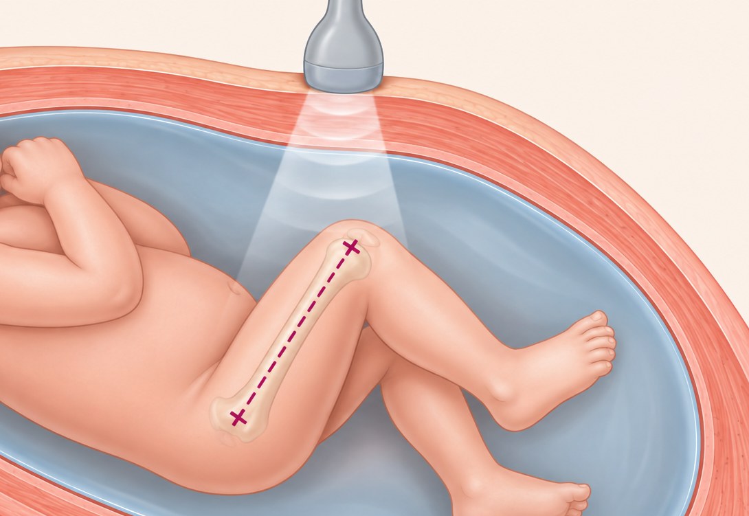
© The Pregnancy Clinic
Why might the femur be short?
There are four main explanations. The scan findings, your earlier screening results and your family background help to work out which is most likely.
1. Early slowing of growth
The long bones can be one of the first measurements to fall behind when the placenta is not supplying the baby as well as it should. This is why a short femur is followed by growth scans later in pregnancy.
2. Chromosomal conditions
A short femur is one of the soft markers for Down’s syndrome. As an isolated marker, with no other associated findings, a short femur does not increase the chance of Down’s syndrome. It matters more when other findings are present.
3. A naturally smaller baby
This is by far the most common reason. Femur length is strongly influenced by the parents’ height, and it also varies with ethnic background. For example, babies of East Asian and South Asian parents often have slightly shorter femurs than the standard charts suggest. These babies are healthy and simply follow their family pattern.
4. Bone conditions (skeletal dysplasia)
Rarely, a short femur is caused by a condition affecting how the bones grow. This is discussed further below.
Is it linked to my baby’s growth?
Yes. This is the most important reason for follow-up. In a large review of more than 3,000 pregnancies with an isolated short femur at 18 to 28 weeks, about 1 in 7 babies were later small for their stage of pregnancy, compared with about 1 in 20 babies with a normal femur length. Babies with a short femur were also slightly more likely to be born early.
If a short femur is linked to a problem with the placenta, there are often other signs that point to this, such as:
- a low PAPP-A level in the combined screening test in the first trimester
- increased resistance in the uterine arteries, which supply blood to the placenta, at the 20-week scan
- a small tummy (abdominal circumference) measurement as well as a short femur.
This is why a specialist fetal medicine review is important. It brings these findings together to work out whether your baby is simply small or whether closer monitoring is needed. Most babies with a short femur grow normally, and growth scans help to identify the smaller number who need extra care, so that the timing of birth can be planned safely if needed.
For further information, read: Uterine Artery Doppler at 20 Weeks: What Does Increased Resistance Mean?
Is it linked to Down’s syndrome?
As an isolated marker, with no other associated findings, a short femur does not increase the chance of Down’s syndrome.
However, a holistic fetal medicine review is essential. This includes a detailed assessment of your baby’s anatomy, a careful check for any other associated markers, and a review of your first-trimester chance of Down’s syndrome from combined screening. If you have had NIPT, this result is also taken into account. If other markers are seen, or you have not had screening, we will discuss whether NIPT or a diagnostic test would be helpful.
For further information, read: Genetic Markers and Soft Markers at the 20-Week Scan
What about skeletal dysplasia?
Skeletal dysplasias are a group of rare conditions that affect how the bones grow. They are much less common than the other explanations. A bone condition is more likely when:
- the femur is very short, well below the usual range
- all the long bones of the arms and legs are short
- the bones look bowed, unusually shaped or appear to have fractures
- the chest looks small, or the head looks large compared with the body.
The most common of these conditions, achondroplasia, often does not show at the 20-week scan. The femur usually measures normally at first and falls behind later, in the third trimester. If the femur is too short, a specialist fetal medicine review should be undertaken to assess your baby’s skeleton in detail, including all the long bones, the spine, the chest, the skull, the hands and the feet.
If a bone condition is suspected, you will be offered a detailed assessment with a fetal medicine specialist and genetic testing, and referral to a team experienced in these conditions.
What happens next?
What is recommended depends on the whole picture, not on the femur measurement alone. As a general guide:
| Scan findings | What it usually means | Usual next step |
|---|---|---|
| Femur short with a small tummy measurement, or reduced blood flow to the placenta | Possible early slowing of growth | Closer growth and Doppler scans |
| Femur short with other soft markers or structural findings | Possible chromosomal condition | Discussion of NIPT or a diagnostic test |
| Femur mildly short, rest of the scan normal | A naturally smaller baby | Growth scan in the third trimester |
| Femur very short, other long bones short or bowed | Possible bone condition | Specialist assessment and genetic testing |
If the femur is mildly short and everything else is normal, a growth scan is usually recommended at around 28 to 32 weeks. If your baby continues to grow well, no further special care is usually needed, and a short femur does not change how or where you give birth.
Frequently asked questions
Did I do something to cause this?
No. A short femur is not caused by anything you have eaten, drunk or done during pregnancy.
Does a short femur mean my baby will be short?
Not necessarily. Many babies with a short femur at 20 weeks grow into children of normal height, often following their parents’ build.
Can the femur catch up?
Yes. Measurements naturally vary between scans, and many femurs that measure short at 20 weeks measure within the normal range at later scans.
Do I need an amniocentesis?
Not usually. When the femur is the only finding and your earlier screening was low chance, a diagnostic test is not normally recommended. It may be discussed if other findings are seen or a bone condition is suspected.
How many growth scans will I need?
Most women are offered one or two growth scans in the third trimester. If your baby’s growth slows, scans are usually repeated every two to three weeks.
Follow-up at The Pregnancy Clinic
At The Pregnancy Clinic in Sevenoaks, Kent, follow-up scans for a short femur are carried out by fetal medicine specialists. Our approach includes:
- careful measurement of all the long bones and a detailed check of your baby’s anatomy
- assessment of your baby’s growth and the blood flow in the placenta and baby
- a review of your earlier screening results and family background
- a clear explanation of the findings on the day
- a written report to share with your NHS maternity team.
Book a follow-up scan
If a short femur was seen at your 20-week scan, a growth and Doppler scan from 28 weeks checks your baby’s growth and the blood flow to the placenta.
Book a growth and Doppler scan →The Pregnancy Clinic provides planned appointments only. If you have pain, bleeding, leaking fluid, reduced baby movements or any other urgent concern, please contact your local NHS maternity unit straight away, or call 999 in an emergency.
Related pregnancy guides
Clinical references
- D’Ambrosio V, Vena F, Marchetti C, et al. Midtrimester isolated short femur and perinatal outcomes: a systematic review and meta-analysis. Acta Obstet Gynecol Scand 2019;98:11–17
- Mailath-Pokorny M, Polterauer S, Worda K, Springer S, Bettelheim D. Isolated short fetal femur length in the second trimester and the association with adverse perinatal outcome. PLoS One 2015;10:e0128820
- Agathokleous M, Chaveeva P, Poon LC, Kosinski P, Nicolaides KH. Meta-analysis of second-trimester markers for trisomy 21. Ultrasound Obstet Gynecol 2013;41:247–61
- Buck Louis GM, Grewal J, Albert PS, et al. Racial/ethnic standards for fetal growth: the NICHD Fetal Growth Studies. Am J Obstet Gynecol 2015;213:449.e1–41
- Friebe-Hoffmann U, Dobravsky L, Friedl TWP, et al. The femur too short? 1373 fetuses with short femur during second-trimester screening. Arch Gynecol Obstet 2022;306:1037–44
This guide provides general information and does not replace individual medical advice. Recommendations may vary according to your scan findings, pregnancy history and clinical circumstances.