
At the 20-week scan, your sonographer checks your baby’s kidneys and bladder. Sometimes the central part of one or both kidneys, where urine collects, looks slightly wider than usual. This is called renal pelvis dilatation, sometimes shortened to RPD, and it may also be described as pyelectasis or mild hydronephrosis.
Renal pelvis dilatation is one of the more common findings at the 20-week scan. In most babies it is mild, causes no problems and settles on its own, either before birth or in the first months of life. A small number of babies need closer monitoring or treatment after birth.
Key points
- Renal pelvis dilatation means the collecting area in the middle of a baby’s kidney is wider than usual.
- It is found in about 1 to 2 in 100 pregnancies and is slightly more common in boys.
- Mild dilatation usually settles on its own and does not affect the baby’s health.
- A follow-up scan later in pregnancy, usually around 32 weeks, checks whether it has improved.
- Some babies are offered a kidney scan after birth, and occasionally antibiotics to prevent urine infections.
- When it is the only finding, it makes very little difference to the chance of Down’s syndrome.
- Larger measurements, both kidneys affected or other findings in the bladder or fluid need a detailed assessment.
What is renal pelvis dilatation?
Each kidney makes urine, which drains into a funnel-shaped space in the middle of the kidney called the renal pelvis. From there, urine passes down a tube called the ureter into the bladder.
If urine drains a little more slowly than usual, the renal pelvis can look slightly wider on the scan. In most babies this is a temporary variation as the urinary system matures. Less often, it is caused by a narrowing where the kidney joins the ureter, urine flowing back up from the bladder (called reflux), or, rarely, a blockage further down.
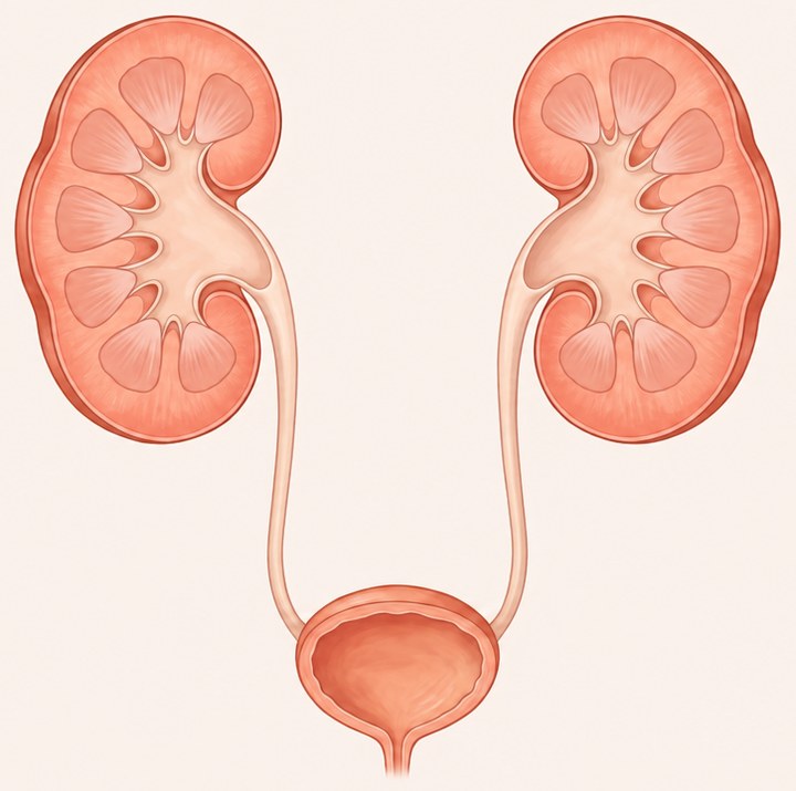 Normal kidneys
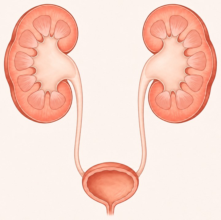 Renal pelvis dilatation
© The Pregnancy Clinic
How is it measured?
The width of the renal pelvis is measured across the kidney, from front to back, in millimetres. This is called the anteroposterior diameter. Both kidneys are measured, and the scan also looks at the rest of the urinary system:
- whether the smaller drainage channels inside the kidney (calyces) are also widened
- the thickness and appearance of the kidney tissue
- whether the ureters can be seen, which usually means they are widened
- the size and wall of the bladder
- the amount of fluid around the baby, which is made mainly from the baby’s urine.
What measurement is normal?
The kidneys and the renal pelvis grow during pregnancy, so the measurement is interpreted according to the stage of pregnancy. Widely used international guidance groups the measurements as follows:
| Stage of pregnancy | Usually normal | Mild dilatation | Needs closer assessment |
|---|---|---|---|
| 16 to 27 weeks | Less than 4 mm | 4 to 7 mm | 7 mm or more |
| 28 weeks or later | Less than 7 mm | 7 to 10 mm | 10 mm or more |
Different hospitals use slightly different cut-offs, and the measurement is always interpreted together with the other findings listed above. A single measurement close to a cut-off is not a diagnosis.
How common is it?
Some widening of the urinary tract is seen in about 1 to 2 in 100 pregnancies. It is slightly more common in boys, and it may affect one kidney or both.
The great majority of babies with mild dilatation have healthy kidneys. In a large review of babies followed up after birth, about 1 in 8 babies with mild dilatation had an underlying kidney or urinary condition, compared with about half of those with moderate dilatation and most of those with severe dilatation. Many of these conditions are minor and improve with time.
Is it linked to Down’s syndrome?
Mild renal pelvis dilatation is one of the soft markers that can be seen at the 20-week scan. When it is the only finding and the rest of the scan is normal, it makes very little difference to the chance of Down’s syndrome.
Your result is interpreted together with your earlier screening. If you had a low-chance combined test or NIPT result in the first trimester, no further testing is usually needed for this reason. If you have not had screening, or other markers are seen, we will discuss whether NIPT would be helpful.
For further information, read: Genetic Markers and Soft Markers at the 20-Week Scan
What happens next in pregnancy?
Most babies with renal pelvis dilatation simply need a follow-up scan later in pregnancy, usually at around 32 weeks, to see whether the measurement has stayed the same, improved or increased.
- If the measurement has settled: no further scans are usually needed for the kidneys.
- If the dilatation persists: your baby will usually be offered a kidney scan after birth, and the paediatric team will be informed.
- If the dilatation is larger, affects both kidneys, or there are changes in the bladder or fluid: you will be offered a detailed assessment with a fetal medicine specialist, and a plan will be made with the paediatric kidney team.
Renal pelvis dilatation does not usually change how or where you give birth.
What happens after birth?
If the dilatation was still present on the later pregnancy scan, your baby will usually be checked by a paediatrician after birth. The plan may include:
- a kidney scan after birth, usually after the first few days, because scans in the first 48 hours can underestimate the measurement while the baby is getting used to feeding
- a small daily dose of antibiotic in some babies, to reduce the chance of a urine infection until the scan results are known
- further tests, only if the scan after birth shows significant dilatation.
Most babies need no treatment. A small number have a condition that needs follow-up by a paediatric kidney specialist, and occasionally surgery is needed to improve drainage.
After birth, it is helpful to look out for signs of a urine infection, such as fever, poor feeding, vomiting or unusual irritability, and to seek medical advice promptly if these occur.
When is it more significant?
Renal pelvis dilatation is more likely to need close follow-up when:
- the measurement is 10 mm or more, or increases between scans
- both kidneys are affected
- the calyces are also widened, or the kidney tissue looks thin or bright
- the ureters can be seen
- the bladder is large or has a thick wall, particularly in a baby boy
- the amount of fluid around the baby is reduced.
These findings do not always mean a serious problem, but they do mean that a detailed scan and specialist advice are recommended.
Frequently asked questions
Did I do something to cause this?
No. Renal pelvis dilatation is not caused by anything you have eaten, drunk or done during pregnancy, and there is nothing you could have done to prevent it.
Can renal pelvis dilatation go away on its own?
Yes. In most babies with mild dilatation, the measurement improves before birth or in the first months of life without any treatment.
Will my baby need surgery?
Most babies do not. Only a small number of babies, usually those with larger or increasing dilatation after birth, need an operation to improve the drainage of urine from the kidney.
Will it affect how I give birth?
Usually not. Renal pelvis dilatation does not normally change the timing or method of delivery. If more significant findings are seen, your care team will discuss whether delivery should be planned in a hospital with specialist paediatric services.
Will my baby need antibiotics?
Some babies are given a small daily dose of antibiotic after birth to reduce the chance of a urine infection, until a kidney scan has been done. Your paediatric team will advise whether this is needed.
Follow-up at The Pregnancy Clinic
At The Pregnancy Clinic in Sevenoaks, Kent, follow-up scans for renal pelvis dilatation are carried out by fetal medicine specialists. Our approach includes:
- a detailed assessment of both kidneys, the ureters, the bladder and the fluid around your baby
- accurate measurement and comparison with your earlier scans
- a review of your earlier screening results, so you have a clear picture of your baby’s chance of chromosomal conditions
- a clear explanation of the findings on the day
- a written report to share with your NHS maternity and paediatric teams.
Book a follow-up scan
If renal pelvis dilatation was seen at your 20-week scan, a growth and Doppler scan from 28 weeks includes a detailed check of your baby’s kidneys.
Book a growth and Doppler scan →The Pregnancy Clinic provides planned appointments only. If you have pain, bleeding, leaking fluid, reduced baby movements or any other urgent concern, please contact your local NHS maternity unit straight away, or call 999 in an emergency.
Related pregnancy guides
Clinical references
- Nguyen HT, Benson CB, Bromley B, et al. Multidisciplinary consensus on the classification of prenatal and postnatal urinary tract dilation (UTD classification system). J Pediatr Urol 2014;10:982–98
- Lee RS, Cendron M, Kinnamon DD, Nguyen HT. Antenatal hydronephrosis as a predictor of postnatal outcome: a meta-analysis. Pediatrics 2006;118:586–93
- Agathokleous M, Chaveeva P, Poon LC, Kosinski P, Nicolaides KH. Meta-analysis of second-trimester markers for trisomy 21. Ultrasound Obstet Gynecol 2013;41:247–61
- Public Health Wales, Antenatal Screening Wales. Renal pelvis dilatation: information for parents
This guide provides general information and does not replace individual medical advice. Recommendations may vary according to your scan findings, pregnancy history and clinical circumstances.