
Being told that you have a subchorionic haematoma during an early pregnancy scan can be worrying, particularly if you have experienced vaginal bleeding.
- Sub
- Under
- Chorion
- A membrane that surrounds the pregnancy sac and contributes to the development of the placenta
- Haematoma
- A clot that forms from collected blood
A subchorionic haematoma is a collection of blood that develops between the membranes surrounding the pregnancy and the wall of the uterus. It is one of the findings that may be identified during an ultrasound examination in women who experience bleeding during early pregnancy.
Most women with a small subchorionic haematoma go on to have a normally developing pregnancy. However, the significance depends on several factors, including the size and location of the haematoma, the gestational age and whether bleeding continues.
Understanding the ultrasound findings can help explain what to expect and whether further monitoring is necessary.
Key points
- During early pregnancy, the developing gestational sac is surrounded by a membrane called the chorion, which contributes to the formation of the placenta.
- A subchorionic haematoma is usually diagnosed during an ultrasound examination.
- The size of a subchorionic haematoma can influence its clinical significance.
- The position of a haematoma may also influence its significance.
What is a subchorionic haematoma?
During early pregnancy, the developing gestational sac is surrounded by a membrane called the chorion, which contributes to the formation of the placenta.
A subchorionic haematoma develops when blood collects between the chorionic membrane and the lining of the uterus. The collection may be small and localised or extend around a larger proportion of the gestational sac. A subchorionic haematoma is sometimes described as a subchorionic bleed or subchorionic haemorrhage.
On ultrasound, it is usually seen as a crescent-shaped or irregular area adjacent to the pregnancy sac. Its appearance can vary depending on whether the blood is fresh or has been present for some time.
Although the finding can be associated with vaginal bleeding, some women have no symptoms, and the haematoma is discovered incidentally during a routine pregnancy scan.
What causes a subchorionic haematoma?
The precise cause is not always known.
A subchorionic haematoma is thought to develop when a small area of the chorionic membrane separates from the underlying uterine lining, allowing blood to collect within the space. This may occur during the early development of the placenta and its attachment to the uterine wall.
In many cases, there is no identifiable reason why the bleeding has occurred. A subchorionic haematoma is not usually caused by normal daily activities, walking, working or routine exercise.
It is important to recognise that the finding does not necessarily indicate that the pregnancy is developing abnormally.
What are the symptoms of a subchorionic haematoma?
The most common symptom is vaginal bleeding. The amount of vaginal bleeding does not always reflect the size of the haematoma seen on ultrasound.
The bleeding may vary considerably between women and can include light spotting, brown discharge, fresh red bleeding or, occasionally, heavier bleeding with small clots.
Brown discharge usually represents older blood that has taken longer to leave the uterus, whereas bright red bleeding generally indicates more recent bleeding. However, the colour of the blood alone cannot determine whether the haematoma is increasing or whether the pregnancy is at risk.
Some women experience intermittent bleeding over several days or weeks as blood from the collection gradually drains through the cervix. Others have no bleeding at all.
Mild abdominal discomfort or cramps may accompany the bleeding, but significant or worsening pain requires medical assessment.
Read more: Bleeding in Early Pregnancy: What Does It Mean?
How is a subchorionic haematoma diagnosed?
A subchorionic haematoma is usually diagnosed during an ultrasound examination.
The clinician assesses the gestational sac, developing embryo, fetal heart activity and the surrounding uterine tissues. A haematoma may appear as a dark or mixed-density area between the pregnancy membranes and the uterine wall.
The examination may also assess:
- The size and extent of the haematoma.
- Its position in relation to the gestational sac and developing placenta.
- Whether the pregnancy is developing appropriately for its gestational age.
- Whether fetal heart activity is present.
- Whether there are other findings that may explain the bleeding.
During the earliest weeks, a transvaginal ultrasound may provide clearer views of the pregnancy and the surrounding tissues. A transvaginal examination is considered safe and does not increase the risk of miscarriage.
Read more: When Can You See a Baby’s Heartbeat on an Early Pregnancy Scan?
Does the size of the haematoma matter?
The size of a subchorionic haematoma can influence its clinical significance.
A small collection adjacent to an otherwise normally developing pregnancy is generally reassuring. Larger collections, particularly those extending around a substantial proportion of the gestational sac, may be associated with an increased risk of pregnancy complications.
However, there is no universally accepted measurement that reliably distinguishes a low-risk haematoma from a high-risk one. The absolute size in millimetres may be less informative than its size relative to the gestational sac or developing placenta.
For example, a collection measuring 20 mm may have a different significance at six weeks compared with 12 weeks, when the pregnancy is considerably larger.
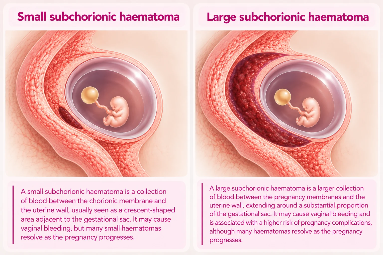
The size of a haematoma should be interpreted in relation to the gestational age, pregnancy development and other ultrasound findings.
Does the location of the haematoma matter?
The position of a haematoma may also influence its significance.
During early pregnancy, collections are commonly seen adjacent to the gestational sac.
As the placenta develops, a collection may be described according to its relationship with the placental edge or the membranes. Some studies suggest that collections situated behind the developing placenta may be associated with greater risk than small collections located at the margins of the gestational sac.
However, these findings are not consistent across all studies, and the location alone cannot reliably predict the outcome. The clinician should consider the overall ultrasound appearance rather than relying on a single measurement or description.
Does a subchorionic haematoma increase the risk of miscarriage?
Research suggests that subchorionic haematomas are associated with an increased risk of miscarriage in some pregnancies.
However, the degree of risk varies considerably between studies and depends on factors such as maternal age, gestational age, haematoma characteristics and the presence of vaginal bleeding.
A small subchorionic haematoma with an appropriately developing embryo and visible heartbeat is usually associated with a favourable pregnancy outcome.
Larger or persistent haematomas may carry a greater risk, but their presence does not mean that miscarriage is inevitable.
The most useful assessment considers the size and location of the collection alongside fetal growth, heart activity and any continuing symptoms.
Read more: Miscarriage in Early Pregnancy: How Likely Is It and Why Does It Happen?
Can a subchorionic haematoma affect pregnancy later on?
Most subchorionic haematomas identified during the first trimester gradually reduce in size or resolve as pregnancy progresses.
However, some studies have reported associations between larger or persistent haematomas and later pregnancy complications, including preterm birth, premature rupture of membranes and placental abruption.
These associations do not mean that a subchorionic haematoma will necessarily cause these complications. The overall evidence is mixed, and many women with a haematoma have an uncomplicated pregnancy.
If a large collection persists into the second trimester, your clinician may recommend additional ultrasound examinations to assess fetal growth, placental development and the appearance of the haematoma.
The need for additional monitoring should be individualised rather than applied routinely to every small haematoma.
How long does a subchorionic haematoma take to disappear?
There is no fixed timeframe.
Some small haematomas resolve within a few weeks, while larger collections may remain visible for considerably longer.
Occasionally, a haematoma may persist into the second trimester. On follow-up ultrasound, the collection may become smaller, change shape or alter in appearance as the blood is gradually broken down and absorbed.
Some women continue to experience brown discharge as older blood leaves the uterus. A haematoma does not necessarily need to disappear completely before the pregnancy can continue normally.
The most reassuring findings are appropriate fetal growth, visible heart activity and a stable or reducing haematoma.
Do I need another ultrasound scan?
Not every subchorionic haematoma requires repeated ultrasound monitoring.
If the collection is small, the pregnancy is developing normally and the bleeding has settled, routine pregnancy care may be sufficient.
A follow-up examination may be recommended when:
- The haematoma is large relative to the gestational sac.
- Bleeding is persistent or recurrent.
- The collection appears to be increasing in size.
- The haematoma is situated close to or behind the developing placenta.
- There are concerns about pregnancy development or other ultrasound findings.
A repeat scan can assess whether the collection is reducing and confirm that the embryo or fetus continues to grow appropriately.
There is no single recommended scanning interval for every woman with a subchorionic haematoma. The timing should depend on the clinical circumstances.
Is there any treatment for a subchorionic haematoma?
There is currently no treatment proven to make a subchorionic haematoma disappear more quickly. Management is usually based on the symptoms, ultrasound findings and overall pregnancy risk.
Bed rest
There is no good evidence that strict bed rest prevents miscarriage or improves outcomes in women with a subchorionic haematoma. Prolonged inactivity may also have disadvantages, including an increased risk of blood clots.
Exercise and sexual intercourse
There is insufficient evidence that routinely avoiding exercise or sexual intercourse improves the outcome of a subchorionic haematoma.
However, when bleeding is active or recurrent, your clinician may recommend temporarily reducing strenuous physical activity or avoiding intercourse based on your individual circumstances. Such advice is precautionary rather than a proven treatment.
Progesterone
Progesterone does not directly treat or remove a subchorionic haematoma. However, NICE recommends vaginal micronised progesterone 400 mg twice daily for women with bleeding in early pregnancy who have had at least one previous miscarriage and have an intrauterine pregnancy confirmed on ultrasound.
If fetal heart activity is confirmed, treatment is continued until 16 completed weeks of pregnancy. This recommendation relates to early pregnancy bleeding and previous miscarriage rather than the presence of a haematoma alone.
Other medication
Aspirin, heparin or other medications should not be started solely to treat a subchorionic haematoma.
If you are already taking aspirin or anticoagulant medication for another medical reason, your clinician should review the indication and the bleeding risk before making any changes. Do not stop prescribed medication without medical advice.
When should I seek urgent medical attention?
If you experience vaginal bleeding during pregnancy, particularly if it is new or increasing, you should contact your GP, midwife or local NHS Early Pregnancy Assessment Service for advice.
Seek urgent medical assessment if you develop heavy bleeding, severe or worsening abdominal pain, significant one-sided pelvic pain, shoulder-tip pain, dizziness or fainting.
These symptoms may indicate significant bleeding or another pregnancy complication.
If you experience collapse, severe pain or heavy bleeding with faintness, call 999 or attend an Emergency Department immediately.
If your pregnancy’s location has not yet been confirmed on ultrasound, it is particularly important not to assume that bleeding is caused by a subchorionic haematoma.
Understanding your subchorionic haematoma
A subchorionic haematoma is a collection of blood adjacent to the developing pregnancy and is one possible cause of bleeding during the first trimester.
Although the finding can be worrying, most small haematomas resolve naturally, and many women continue to have a healthy pregnancy.
The significance depends on the size and location of the collection, whether bleeding persists and, most importantly, how the pregnancy is developing.
A reassuring ultrasound showing appropriate fetal growth and heart activity is generally more informative than the presence of a small haematoma alone.
Where additional monitoring is appropriate, follow-up ultrasound can help assess the haematoma and provide reassurance about continuing pregnancy development.
Related pregnancy guides
Frequently asked questions
What is a subchorionic haematoma?
During early pregnancy, the developing gestational sac is surrounded by a membrane called the chorion, which contributes to the formation of the placenta.
What causes a subchorionic haematoma?
The precise cause is not always known. A subchorionic haematoma is thought to develop when a small area of the chorionic membrane separates from the underlying uterine lining, allowing blood to collect within the space.
What are the symptoms of a subchorionic haematoma?
The most common symptom is vaginal bleeding. The amount of vaginal bleeding does not always reflect the size of the haematoma seen on ultrasound.
How is a subchorionic haematoma diagnosed?
A subchorionic haematoma is usually diagnosed during an ultrasound examination.
Does the size of the haematoma matter?
The size of a subchorionic haematoma can influence its clinical significance.
Does the location of the haematoma matter?
The position of a haematoma may also influence its significance. During early pregnancy, collections are commonly seen adjacent to the gestational sac.
Early pregnancy care at The Pregnancy Clinic
At The Pregnancy Clinic, we provide consultant-led early pregnancy ultrasound examinations, including assessment of vaginal bleeding and subchorionic haematomas.
Our approach is to evaluate the size and location of the haematoma, assess pregnancy development and explain the significance of the findings in the context of your individual circumstances.
Where appropriate, we can arrange follow-up ultrasound examinations and provide guidance on activity, medication and further pregnancy care.
Book an early pregnancy consultation → Book an early pregnancy scan →The Pregnancy Clinic is not an emergency service. If you experience severe pain, heavy bleeding, fainting or other acute symptoms, please seek urgent NHS assessment.