
An ectopic pregnancy occurs when a pregnancy develops outside the normal cavity of the uterus. It affects approximately 1 in 90 pregnancies in the UK.
Being told that you may have an ectopic pregnancy can be frightening, particularly when the diagnosis is uncertain or you have experienced bleeding or abdominal pain.
An ectopic pregnancy cannot develop normally and may become life-threatening if it causes internal bleeding. Early diagnosis and appropriate treatment are therefore essential.
However, not every woman with bleeding or abdominal pain has an ectopic pregnancy, and an ultrasound performed very early may not immediately establish where the pregnancy is located.
Key points
- An ectopic pregnancy occurs when a fertilised egg implants and develops outside the normal cavity of the uterus.
- The uterus is specifically designed to accommodate and support a growing pregnancy.
- Symptoms most commonly develop between approximately four and twelve weeks of pregnancy, although they may occur earlier or later.
- An ectopic pregnancy can occur in any woman, including those without identifiable risk factors.
What is an ectopic pregnancy?
An ectopic pregnancy occurs when a fertilised egg implants and develops outside the normal cavity of the uterus.
What are the different types of ectopic pregnancy?
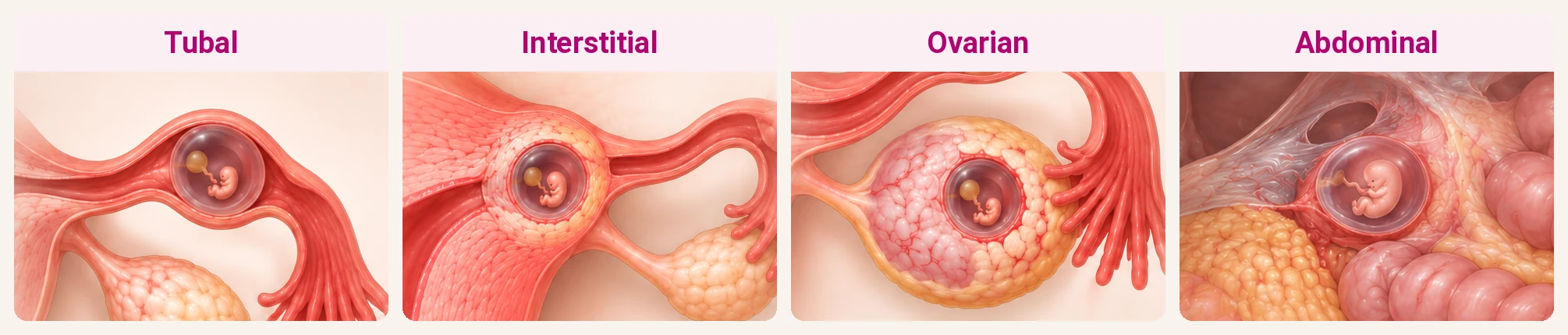
Ectopic pregnancies are classified according to where the pregnancy implants. The different types, listed approximately in order of frequency, are:
- Tubal ectopic pregnancy. This is the most common type of ectopic pregnancy. The pregnancy implants within a fallopian tube, most commonly in the ampullary portion of the tube.
- Interstitial ectopic pregnancy. The pregnancy implants within the interstitial portion of the fallopian tube, where the tube passes through the muscular wall of the uterus.
- Ovarian ectopic pregnancy. The pregnancy implants within the ovary.
- Caesarean scar ectopic pregnancy. The pregnancy implants within the scar of a previous Caesarean section. This is an uncommon but increasingly recognised type of ectopic pregnancy.
- Cervical ectopic pregnancy. The pregnancy implants within the cervix rather than the uterine cavity. This is a very rare type of ectopic pregnancy.
- Abdominal ectopic pregnancy. The pregnancy implants within the abdominal cavity, outside the uterus and fallopian tubes. This is extremely rare.
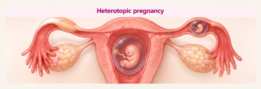
Heterotopic pregnancy
A heterotopic pregnancy is a separate condition in which two pregnancies develop simultaneously: one within the normal uterine cavity and another outside it, most commonly within a fallopian tube.
Heterotopic pregnancy is extremely rare following natural conception but is more common following assisted reproductive treatment, including IVF.
Why is an ectopic pregnancy a cause for concern?
The uterus is specifically designed to accommodate and support a growing pregnancy. Its muscular walls can expand considerably as the pregnancy develops, while the uterine lining and placenta provide the environment necessary for fetal growth.
In the most common type of ectopic pregnancy, a tubal ectopic pregnancy, the pregnancy implants within a fallopian tube. As the pregnancy grows, the fallopian tube gradually becomes distended. Unlike the uterus, the fallopian tube is not designed to accommodate an enlarging pregnancy, and its tissues have a limited capacity to stretch.
As the pregnancy continues to develop, the tissues of the fallopian tube may eventually be unable to accommodate further growth. This can cause the tube to tear or rupture, resulting in a ruptured ectopic pregnancy. Rupture can cause significant bleeding into the abdominal cavity, sometimes leading to rapid blood loss, circulatory collapse and a life-threatening emergency requiring immediate surgical treatment.
Importantly, rupture can occur even when the pregnancy is relatively small or at an early gestational age.
Similarly, tissues outside the normal uterine cavity are not biologically designed to support a growing and advancing pregnancy. Ectopic pregnancies in other locations may therefore cause serious complications, particularly bleeding, as the pregnancy develops and invades surrounding tissues.
Some ectopic pregnancies stop developing spontaneously, but others continue to grow and may cause significant complications.
An ectopic pregnancy is therefore a cause for concern regardless of its location, and early diagnosis, appropriate monitoring and timely treatment are essential to prevent potentially life-threatening complications.
What are the symptoms of an ectopic pregnancy?
Symptoms most commonly develop between approximately four and twelve weeks of pregnancy, although they may occur earlier or later. The most common symptoms include vaginal bleeding and abdominal or pelvic pain.
Vaginal bleeding
Bleeding may be light or heavy and can appear as spotting, brown discharge or fresh red blood. Some women experience irregular bleeding that differs from their usual menstrual period.
Abdominal or pelvic pain
Pain may occur on one side of the lower abdomen, although it can sometimes be more generalised. It may develop gradually, be intermittent or become progressively more severe.
Shoulder-tip pain
Pain at the tip of the shoulder can occur when internal bleeding irritates the diaphragm. This is an important warning symptom and requires urgent assessment.
Dizziness, fainting or collapse
These symptoms may indicate significant internal bleeding and require immediate emergency medical attention.
Some women have few or no symptoms during the earliest stages, and an ectopic pregnancy may be suspected during an ultrasound examination before significant pain or bleeding develops.
The severity of symptoms does not always reflect the size of the ectopic pregnancy.
Read more: Bleeding in Early Pregnancy: What Does It Mean?
Who is at increased risk of ectopic pregnancy?
An ectopic pregnancy can occur in any woman, including those without identifiable risk factors. However, certain circumstances increase the likelihood. These include:
- A previous ectopic pregnancy.
- Previous pelvic infection, particularly involving the fallopian tubes.
- Previous surgery involving the fallopian tubes.
- Fertility treatment, including IVF.
- Smoking.
- Pregnancy occurring with an intrauterine contraceptive device in place.
Most pregnancies are prevented by an intrauterine contraceptive device. However, if pregnancy does occur, the possibility of an ectopic pregnancy requires particular consideration.
Many women diagnosed with an ectopic pregnancy have no known risk factors. The absence of risk factors should therefore never be used to exclude the diagnosis.
How is ectopic pregnancy diagnosed?
Diagnosis usually involves a combination of clinical assessment, transvaginal ultrasound and, when necessary, serial blood tests measuring the pregnancy hormone β-hCG.
The clinician will consider your symptoms, medical history, pregnancy dates and ultrasound findings.
Transvaginal ultrasound is the most important imaging investigation for suspected ectopic pregnancy.
During the examination, the clinician assesses the uterine cavity, both ovaries, the regions surrounding the fallopian tubes and the pelvis for any evidence of internal bleeding. Sometimes an ectopic pregnancy can be identified directly. In other circumstances, the ultrasound findings may be uncertain, particularly when the pregnancy is very early.
What does an ectopic pregnancy look like on ultrasound?
The appearance depends on the location and stage of development.
An ectopic pregnancy may be identified as a gestational sac outside the uterus containing a yolk sac or embryo. Occasionally, heart activity may also be visible. These findings provide strong evidence of an ectopic pregnancy. More commonly, ultrasound may demonstrate an abnormal mass adjacent to an ovary or within the region of a fallopian tube.
The clinician may also identify fluid within the pelvis, which can sometimes represent blood.
The ultrasound findings must be interpreted carefully to avoid confusing an early intrauterine pregnancy, ovarian corpus luteum or other pelvic structures with an ectopic pregnancy.
What if nothing is visible on ultrasound?
Sometimes a woman has a positive pregnancy test, but ultrasound does not demonstrate a definite pregnancy either inside or outside the uterus. This is called a pregnancy of unknown location (PUL).
There are several possible explanations. The pregnancy may be developing normally within the uterus but be too early to see. Alternatively, the pregnancy may have stopped developing at a very early stage, or it may be an ectopic pregnancy that is not yet visible.
A pregnancy of unknown location is therefore not the same as a confirmed ectopic pregnancy. However, an ectopic pregnancy cannot be excluded until further assessment establishes the location or confirms that the pregnancy has resolved.
Read more: Pregnancy of Unknown Location: What Happens Next?
Can hCG blood tests diagnose an ectopic pregnancy?
Human chorionic gonadotrophin (β-hCG) is the pregnancy hormone measured in blood tests.
During early pregnancy, hCG levels generally increase as the pregnancy develops. When ultrasound cannot establish the pregnancy’s location, two blood tests taken approximately 48 hours apart may help guide further assessment. In some ectopic pregnancies, hCG rises more slowly than expected.
However, there is no particular hCG level or pattern of increase that can reliably diagnose or exclude an ectopic pregnancy.
Some ectopic pregnancies demonstrate substantial increases in hCG, while others may show falling hormone levels. NICE recommends using serial hCG measurements to guide the management of a pregnancy of unknown location, rather than to determine where the pregnancy is located. A falling hCG level does not completely eliminate the risk of complications.
Clinical symptoms and ultrasound findings remain essential, regardless of the hCG result.
Read more: Understanding hCG Levels in Early Pregnancy
Can an ectopic pregnancy be mistaken for a normal early pregnancy?
During the earliest weeks, it may be difficult to distinguish a very early intrauterine pregnancy from a pregnancy of unknown location.
A small fluid collection within the uterus does not necessarily establish that a pregnancy has implanted normally. Identifying a gestational sac containing a yolk sac or embryo within the uterine cavity provides much stronger evidence of an intrauterine pregnancy.
Once a definite intrauterine pregnancy has been identified, an ectopic pregnancy is very unlikely in a spontaneous singleton pregnancy. However, the rare possibility of a simultaneous intrauterine and ectopic pregnancy should be considered when there are concerning symptoms, particularly following IVF.
Read more: What Can You See on a Pregnancy Scan at 5, 6, 7 and 8 Weeks?
How is an ectopic pregnancy treated?
Treatment depends on the woman’s symptoms, clinical stability, ultrasound findings, hCG levels and the characteristics of the ectopic pregnancy. The three main approaches are expectant management, medical treatment and surgery.
Expectant management
Some small ectopic pregnancies resolve naturally without active treatment. This approach may be appropriate in carefully selected women who are clinically stable, have no significant pain and meet specific ultrasound and hCG criteria. Regular blood tests and reliable follow-up are essential until the pregnancy has resolved.
Medical treatment
Methotrexate is a medication that stops the pregnancy tissue from continuing to develop. It may be suitable for selected women with an unruptured ectopic pregnancy who are clinically stable and meet the necessary treatment criteria. Follow-up blood tests are required to confirm that hCG levels are falling appropriately.
Methotrexate is not suitable for every ectopic pregnancy, and pregnancy must be avoided for a period after treatment according to specialist advice.
Surgical treatment
Surgery may be necessary when there is significant pain, internal bleeding, suspected rupture or when medical or expectant management is unsuitable.
Laparoscopic surgery is commonly used when the woman is clinically stable. Depending on the circumstances, the affected fallopian tube may be removed or an attempt made to remove the ectopic pregnancy while preserving the tube. Emergency surgery may be required if an ectopic pregnancy ruptures.
The most appropriate treatment is determined by the clinical findings and should be discussed with the specialist gynaecology team.
Can I have a normal pregnancy after an ectopic pregnancy?
Many women who have experienced an ectopic pregnancy subsequently have a successful intrauterine pregnancy.
The likelihood depends partly on the condition of the remaining fallopian tube, any underlying tubal disease and whether fertility treatment is required. However, the risk of another ectopic pregnancy is higher after a previous ectopic pregnancy. RCOG guidance estimates the recurrence risk at approximately 7–10%.
For this reason, women with a previous ectopic pregnancy are usually advised to contact an Early Pregnancy Assessment Service when they next become pregnant. An early ultrasound, often at approximately six to eight weeks, can help confirm that the pregnancy is developing within the uterus.
Read more: When Should I Have My First Pregnancy Scan?
When should I seek emergency medical attention?
Ectopic pregnancy can cause serious internal bleeding, sometimes before a definite diagnosis has been established. You should seek urgent medical assessment if you have a positive pregnancy test and develop abdominal pain, vaginal bleeding or other concerning symptoms.
Particularly important warning signs include:
- Sudden, severe or worsening abdominal pain.
- Significant pain on one side of the pelvis.
- Shoulder-tip pain.
- Dizziness, fainting or collapse.
- Marked weakness or symptoms suggesting significant blood loss.
If you experience severe abdominal pain, collapse, fainting or symptoms of internal bleeding, call 999 or attend an Emergency Department immediately.
Do not wait for a scheduled ultrasound or blood test if your symptoms change. An ectopic pregnancy may rupture even when hCG levels are relatively low or falling.
Understanding ectopic pregnancy
Ectopic pregnancy occurs when a pregnancy implants outside the normal uterine cavity, most commonly within a fallopian tube.
Although it is relatively uncommon, early recognition is important because an ectopic pregnancy can cause significant internal bleeding.
Transvaginal ultrasound is central to diagnosis, while serial hCG measurements can help guide follow-up when the location remains uncertain.
The most important principle is that an ectopic pregnancy must be considered whenever a woman has a positive pregnancy test and concerning symptoms, particularly when an intrauterine pregnancy has not yet been confirmed.
Early assessment, appropriate follow-up and timely treatment help reduce the risk of serious complications.
Related pregnancy guides
Frequently asked questions
What is an ectopic pregnancy?
An ectopic pregnancy occurs when a fertilised egg implants and develops outside the normal cavity of the uterus.
Why is an ectopic pregnancy a cause for concern?
The uterus is specifically designed to accommodate and support a growing pregnancy. Its muscular walls can expand considerably as the pregnancy develops, while the uterine lining and placenta provide the environment necessary for fetal growth.
What are the symptoms of an ectopic pregnancy?
Symptoms most commonly develop between approximately four and twelve weeks of pregnancy, although they may occur earlier or later. The most common symptoms include vaginal bleeding and abdominal or pelvic pain.
Who is at increased risk of ectopic pregnancy?
An ectopic pregnancy can occur in any woman, including those without identifiable risk factors. These include a previous ectopic pregnancy; previous pelvic infection, particularly involving the fallopian tubes; previous surgery involving the fallopian tubes; fertility treatment, including IVF.
How is ectopic pregnancy diagnosed?
Diagnosis usually involves a combination of clinical assessment, transvaginal ultrasound and, when necessary, serial blood tests measuring the pregnancy hormone β-hCG.
What does an ectopic pregnancy look like on ultrasound?
The appearance depends on the location and stage of development. An ectopic pregnancy may be identified as a gestational sac outside the uterus containing a yolk sac or embryo.
Early pregnancy care at The Pregnancy Clinic
At The Pregnancy Clinic, we provide consultant-led early pregnancy ultrasound examinations, including assessment when the location or development of a pregnancy is uncertain.
Our approach is to evaluate the ultrasound findings carefully, explain their significance and provide clear recommendations for further investigation or follow-up.
Where an ectopic pregnancy is suspected, timely referral to an appropriate NHS Early Pregnancy Assessment Service or emergency gynaecology team is essential.
Book an early pregnancy consultation → Book an early pregnancy scan →The Pregnancy Clinic is not an emergency service. If you have symptoms suggestive of an ectopic pregnancy, particularly severe abdominal pain, shoulder-tip pain, dizziness or fainting, seek urgent NHS assessment rather than waiting for a private appointment.