Menu
Close
Menu
Close
Growth & Doppler Scan
Expert evaluation of fetal growth, placental function and wellbeing to support safe and confident planning for birth
The Growth and Doppler scan will assess how your baby is growing and developing in the later stages of pregnancy.
Using Doppler ultrasound, we also evaluate blood flow in the umbilical cord and baby’s brain, providing important information about placental function and overall fetal well-being.
A late third-trimester assessment, usually around 36 weeks, helps guide planning for birth by assessing growth, baby’s position and placental health.
Book your consultant-led growth scan today for expert assessment, reassurance and high-quality pregnancy care.

FAQs
What are the objectives of the third-trimester growth and wellbeing scan?
The third-trimester Growth and Doppler scan is designed to assess your baby’s growth, wellbeing and placental function during later stages of the pregnancy. The key objectives of the scan include:
- Estimating fetal weight and assessing growth over time
- Evaluating placental blood flow using Doppler ultrasound
- Measuring amniotic fluid volume
- Reassessing fetal anatomy and development, where appropriate
- Confirming fetal presentation (head-down or breech), particularly in late pregnancy
- Reviewing placental location
This scan plays an important role in monitoring your baby’s health during the final stages of pregnancy and supports safe, individualised planning for birth.
What determines a baby’s growth potential?
A baby’s growth potential is influenced mainly by two factors:
- Parental constitution and genetics, which contribute to baby’s expected size
- Placental health and function, which affects transfer of oxygen and nutrients
At the time of the 20-week anomaly scan, most babies are of similar size regardless of parental build. For this reason, estimated weight at 20 weeks does not reliably predict birthweight. Growth assessments in the third trimester, particularly closer to term, provides a much more accurate reflection of growth potential.
When placental function is reduced, growth may be limited, leading to a smaller baby. In contrast, some pregnancies – such as those affected by diabetes – may be associated with increased growth.
Why is it important to assess fetal growth in the third trimester?
Monitoring fetal growth in the third trimester helps identify babies who are smaller or larger than expected for gestational age. Significant deviations from expected growth may be associated with an increased likelihood of complications around the time of birth.
Babies who are significantly small or large are more likely to require closer monitoring. Early identification allows appropriate surveillance, timely referral where needed and personalised plan for ongoing care and delivery.
What if my baby is small or large for gestational age?
Most babies who are small (<10th centile) or large (>90th centile) for gestational age are healthy and have good outcomes.
- Many small babies are constitutionally small and require reassurance rather than intervention
- Babies who are smaller may have reduced natural reserves and may benefit from additional monitoring to understand the underlying cause and guide delivery planning
- Babies with more significant growth restriction may require specialist input and a tailored management plan
Similarly, most larger babies are healthy. In pregnancies complicated by diabetes, closer monitoring is recommended. Where babies are large without diabetes, discussing delivery options helps support informed decision-making.
Why is assessment of amniotic fluid in third trimester important?
Amniotic fluid reflects fetal wellbeing and placental function.
- Normal fluid levels are generally reassuring
- Reduced fluid may indicate placental insufficiency or leaking of waters
- Increased fluid is often a normal variation but may occasionally be associated with maternal or fetal conditions such as diabetes, infection or birth defects
Any variation in amniotic fluid volume is interpreted alongside other scan findings and the overall clinical picture.
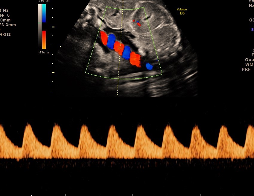
What are Doppler tests and why are they important?
Doppler ultrasound is a specialised technique used to assess blood flow in key fetal and placental blood vessels. During growth scans, Doppler assessment helps evaluate placental function and how baby is adapting to the pregnancy. Commonly assessed vessels include:
- Umbilical artery, reflecting resistance to blood flow across the placenta
- Middle cerebral artery (brain), which can indicate fetal adaptation to reduced oxygen supply
- Ductus venosus (near the heart), assessed in selected cases to reflect fetal heart function
Doppler findings are particularly valuable in the third trimester and may influence monitoring strategies and timing of delivery.
When should baby’s position be checked in the third trimester?
Fetal position changes frequently throughout the pregnancy.
- Breech presentation is relatively common earlier in third trimester
- By 36 weeks, most babies have turned head down
If a baby remains breech at around 36 weeks, this scan provides an opportunity to discuss management options and plan appropriately for delivery.
When should growth scans be performed in the third trimester?
Growth scans can be performed from 26–28 weeks onwards.
- In pregnancies with additional risk factors, scans may be recommended every 2–3 weeks
- In pregnancies without identified risk factors, scans can be performed every 4 weeks
If only one third-trimester scan is undertaken, evidence supports performing it between 35 and 37 weeks, when it provides the most clinically useful information.
I had a normal anomaly scan at 20 weeks – do I need another scan at 36 weeks?
Yes. While the 20-week anomaly scan is the optimal time to assess fetal anatomy, baby’s development continues throughout pregnancy.
Some conditions only become apparent in the third trimester and may not be visible earlier, even after a detailed and expertly performed anomaly scan. A 36-week assessment helps identify findings that may influence newborn care or postnatal follow up.
Most late-presenting findings are mild and manageable, and early identification allows appropriate planning and reassurance.
Difference between NHS, High-street providers and our Consultant-led care pathway for Growth scans?
| Feature |
NHS Care Pathway |
High-Street Scan Providers |
Our Consultant-Led Care Pathway |
|---|---|---|---|
|
Primary purpose |
Not routine care, selective |
Reassurance imaging |
Comprehensive medical assessment |
|
Who performs the scan |
Sonographer |
Sonographer |
Consultant in Fetal Medicine |
|
Appointment duration |
Short, standardised |
Variable |
Longer, unhurried consultation |
|
Assessment of fetal growth |
✅ Standard biometry |
Limited |
✅ Detailed and contextual |
|
Interpretation of growth centiles |
✅ Variable |
❌ |
✅ Consultant interpretation |
|
Serial growth assessment |
✅ Selective cases |
❌ |
✅ Routinely offered and individualised |
|
Umbilical artery Doppler |
✅ When indicated |
❌ |
✅ Routinely assessed |
|
Middle cerebral artery (MCA) Doppler |
Limited |
❌ |
✅ Routinely assessed |
|
Assessment of placental function |
Limited |
❌ |
✅ Comprehensive |
|
Amniotic fluid assessment |
✅ |
Limited |
✅ Detailed |
|
Assessment of fetal wellbeing |
Limited |
❌ |
✅ Comprehensive and full assessment |
|
Immediate clinical interpretation |
Limited |
❌ |
✅ Consultant-led discussion |
|
Continuity of care |
Variable |
❌ |
✅ Consistent consultant oversight |
|
Suitable as a medical assessment |
✅ |
❌ |
✅ |
|
Commonly used to complement NHS care |
|
Occasionally |
✅ Very commonly |