Menu
Close
Menu
Close
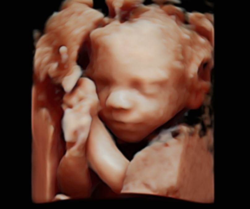
Anomaly Scan
Detailed fetal anatomy, placental assessment and Doppler screening—delivered within a consultant-led care pathway
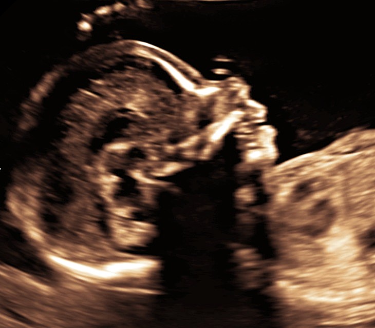
The 20-week anomaly scan is a key milestone in pregnancy and the next major assessment after the nuchal scan. At this stage, your baby’s organs and structures are sufficiently developed to allow a detailed evaluation of growth and anatomy, helping to identify or rule out major structural conditions.
A common misconception is that the anomaly scan focuses only on the baby. At The Pregnancy Clinic, we undertake a detailed review of the placenta and umbilical cord, including screening for placenta and vasa praevia. We perform uterine artery Doppler assessment, which provides information about placental function and helps assess the risk of pregnancy complications such as preeclampsia and fetal growth restriction.
Book your consultant-led anomaly scan today for expert assessment, reassurance and high-quality pregnancy care.

FAQs
What are the objectives of the advanced anomaly scan?
The advanced anomaly scan is a detailed assessment of both the baby and the placenta. Its key objectives include:
- Assessment of the baby’s growth and wellbeing
- A detailed examination of fetal anatomy
- Evaluation of ultrasound markers associated with genetic conditions
- Measurement of amniotic fluid volume
- Assessment of placental location and its relationship to the cervix (neck of the womb)
- In women with a previous caesarean section, assessment of the placenta–scar relationship to exclude abnormal placental attachment
- Evaluation of umbilical cord insertion and screening for vasa praevia
- Uterine artery Doppler assessment to evaluate placental function
This scan plays a crucial role in identifying conditions that may benefit from closer monitoring or early intervention.
Does a normal anomaly scan rule out all birth defects?
Absolutely not. From a parent’s perspective, the key question is not simply whether the baby’s anatomy appears normal, but whether the pregnancy as a whole is healthy and likely to result in a safe, healthy outcome at birth.
Assessment of fetal anatomy and exclusion of structural abnormalities is important, but it represents only one component of a comprehensive mid-pregnancy evaluation. Many conditions that significantly affect pregnancy outcome are not related to fetal anatomy and will be missed if the scan is limited to a purely anatomical checklist.
A high-quality anomaly scan should also include careful assessment of the placenta—its location, appearance, and features suggestive of placental dysfunction—because a healthy pregnancy requires not only a healthy baby, but also a healthy placenta. Similarly, evaluation of the umbilical cord is essential, as cord abnormalities can have serious implications for the baby, for example in conditions such as vasa praevia. The 20-week scan also provides a valuable opportunity to assess the risk of preterm birth through cervical length measurement.
The term “anomaly scan” is unfortunately often interpreted too literally, leading many providers to focus almost exclusively on fetal anatomy. At The Pregnancy Clinic, the advanced anomaly scan is therefore designed as a comprehensive, holistic assessment of the pregnancy—integrating fetal anatomy with placental health, cord assessment, and preterm birth risk—so that families receive meaningful reassurance about the overall health and wellbeing of the pregnancy, not just a structural check of the baby.
What are genetic or soft markers?
Genetic or “soft” markers are subtle ultrasound findings seen during the detailed (anomaly) scan. On their own, they are not structural abnormalities and usually represent minor variations from what is considered a typical anatomical appearance.
In most cases, these markers have no impact on the baby’s health and simply reflect normal variation in development. However, some soft markers can be statistically associated with certain genetic or chromosomal conditions—particularly if more than one marker is present or if there are other risk factors.
It is important that these markers are carefully assessed during the detailed ultrasound scan. When no soft markers are seen, and the baby’s anatomy appears normal, this is very reassuring and significantly reduces the likelihood of an underlying genetic concern.
If a soft marker is identified, this does not automatically mean that there is a problem. In such situations, a discussion with a fetal medicine specialist is helpful to explain the finding in context—taking into account the rest of the scan, previous screening results, and individual risk factors—and to advise whether any further tests or follow-up are needed.
Importantly, soft markers are not uncommon, and in the vast majority of pregnancies they are of no clinical significance, representing normal variations in fetal development rather than evidence of disease.
Does a normal anomaly scan rule out all birth defects?
The accuracy of an anomaly scan depends not only on the quality of the ultrasound equipment, but—crucially—on the skill and expertise of the person performing and interpreting the scan. Even in expert hands, a detailed mid-pregnancy anomaly scan can identify most, but not all, fetal abnormalities.
This is because fetal development is still ongoing at 20 weeks. While many organs are well formed by the second trimester, some continue to develop, mature, or change in appearance and function later in pregnancy. As a result, certain conditions may only become apparent in the third trimester, despite a thorough and normal mid-pregnancy scan.
For this reason, a comprehensive approach to pregnancy care does not rely on a single scan alone. Re-assessment of fetal anatomy and wellbeing later in pregnancy provides additional reassurance for most parents and reduces the risk of unexpected findings after birth. Importantly, it also allows conditions that do emerge later to be identified early enough for appropriate planning and timely management, both during pregnancy and after delivery.
In summary, a normal anomaly scan is an excellent and important milestone, but it should be seen as part of an ongoing, expert-led assessment of fetal health throughout pregnancy rather than a guarantee that every possible condition has been excluded.
What is the importance of amniotic fluid assessment?
Amniotic fluid is primarily made up of fetal urine and circulates continuously through the baby’s body. It is produced by the baby’s kidneys, swallowed, and then absorbed through the intestines.
- A normal amniotic fluid volume suggests normal kidney function and overall fetal wellbeing
- Reduced fluid levels may indicate placental insufficiency or problems with fetal kidney function
- Excessive fluid can also be associated with certain fetal or maternal conditions
Assessing amniotic fluid provides important information about both the baby’s health and placental function.
What is importance of checking location of placenta in the womb?
The placenta (afterbirth) is a pregnancy-specific organ that connects the baby’s circulation to the mother’s. It can be attached to the front (anterior), back (posterior), or sides of the womb.
At the anomaly scan, particular attention is paid to the distance between the placental edge and the cervix:
- If the placental edge lies within 20 mm of the cervix at 20 weeks, it is described as a low-lying placenta
- This is relatively common and in around 90% of cases, the placenta moves upwards as the womb grows
- If the placenta remains within 20 mm of, or covers, the cervix later in pregnancy, this is called placenta praevia
Placenta praevia carries a risk of bleeding during labour and usually requires delivery by caesarean section. At The Pregnancy Clinic, we routinely check for this as early identification allows safe planning and pregnancy monitoring.
What is importance of checking umbilical cord insertion and what is vasa praevia?
The umbilical cord normally inserts into the central portion of the placenta. In some pregnancies, it may insert:
- At the edge of the placenta (marginal insertion), or
- Into the membranes outside the placental tissue (velamentous cord insertion)
When a velamentous cord insertion occurs in the lower part of the womb, exposed fetal blood vessels may cross near the cervix. This condition is known as vasa praevia.
- These unprotected vessels are at risk of rupture during labour or when membranes rupture
- Undiagnosed vasa praevia is associated with serious risk to the baby
- When identified antenatally, careful monitoring and planned delivery lead to excellent outcomes
At The Pregnancy clinic, this is part of the detailed assessment as a focused ultrasound assessment of cord insertion can reliably confirm or exclude this condition.
What is an abnormally invasive placenta or placenta accreta spectrum?
An abnormally invasive placenta occurs when the placenta attaches too deeply into the muscle of the womb and does not separate normally after birth.
- This can result in severe, life-threatening bleeding at delivery
- The strongest risk factor is a previous caesarean section, particularly when the placenta lies over the scar
At the anomaly scan, special attention is given to the relationship between the placenta and any previous uterine scar. If abnormal attachment is suspected, early referral to a specialist centre is essential to ensure safe pregnancy and delivery planning.
What is uterine artery Doppler and how does it help to assess placental function?
Doppler ultrasound is a specialised technique used to assess blood flow within blood vessels.
- The uterine arteries supply blood from the mother to the placenta
- Doppler assessment measures the resistance and pattern of blood flow in these vessels
Increased resistance is associated with placental insufficiency, which can increase the risk of:
- Pre-eclampsia
- Fetal growth restriction
At The Pregnancy Clinic, uterine artery Doppler assessment is routinely included in the anomaly scan to help evaluate placental health and guide personalised pregnancy care.
Difference between NHS, High-street providers and our Consultant-led care pathway for Anomaly scans?
| Feature |
NHS Care Pathway |
High-Street Scan Providers |
Our Consultant-Led Care Pathway |
|---|---|---|---|
|
Primary purpose |
National programme |
Reassurance imaging |
Comprehensive medical assessment |
|
Who performs the scan |
Sonographer |
Sonographer |
Consultant in Fetal Medicine |
|
Appointment duration |
Short, standardised |
Variable |
Longer, unhurried consultation |
|
Assessment of fetal growth |
✅ |
Limited |
✅ Detailed and contextual |
|
Detailed fetal anatomy review |
✅ Standard protocol |
❌ |
✅ Advanced, systematic review |
|
Assessment of genetic markers |
Limited |
❌ |
✅ Expert evaluation |
|
Amniotic fluid assessment |
✅ |
Limited |
✅ Comprehensive |
|
Placental location and cervix relationship |
✅ |
❌ |
✅ Detailed |
|
Assessment of placenta over CS scar |
Limited |
❌ |
✅ Specialist assessment |
|
Umbilical cord insertion |
Not routine |
❌ |
✅ Routinely assessed |
|
Screening for vasa praevia |
Not routine |
❌ |
✅ Routinely screened |
|
Uterine artery Doppler |
Not routine |
❌ |
✅ Included |
|
Screening for placental insufficiency |
Limited |
❌ |
✅ Proactive & detailed |
|
Screening for preterm birth risk |
Not routine |
❌ |
✅ Included |
|
Immediate clinical interpretation |
Limited |
❌ |
✅ Consultant-led discussion |
|
Continuity of care |
Variable |
❌ |
✅ Consistent consultant oversight |
|
Suitable as a medical assessment |
✅ |
❌ |
✅ |
|
Commonly used to complement NHS care |
|
Occasionally |
✅ Very commonly |